
 Belgium/Luxembourg (English)




Budd-Chiari Syndrome and Dioctophyma renale
Identification and history
- Name: Brujo
- Report and medical history: feline, Common European, male, 4-moths-old.
Brujo is a 4-month-old Argentine rescue cat (Common European) presented with persistent abdominal effusion. Tests for common viral diseases were negative, and the Rivalta test was also negative. Physical examination revealed abdominal distension without any other remarkable clinical signs. Bloodwork showed anisocytosis, polychromasia, and increased ALT and AST activities. Cytological evaluation of the effusion was consistent with a modified transudate, likely associated with a subacute inflammatory process.
Diagnostics
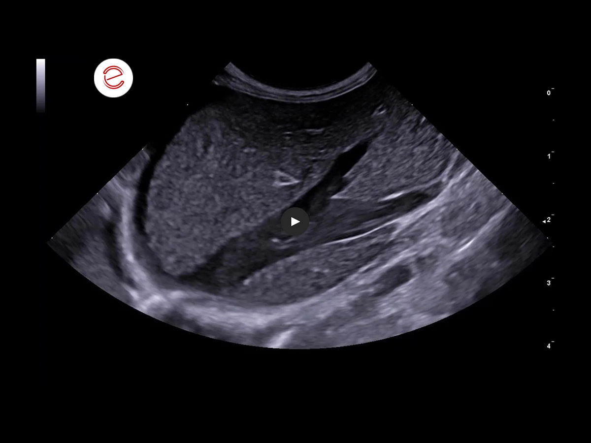

Longitudinal view of the hepatic and caval veins obtained from a right lateral coronal view.
The caudal vena cava and hepatic veins are distended, with spontaneous venous flow that does not adequately progress toward the thorax.
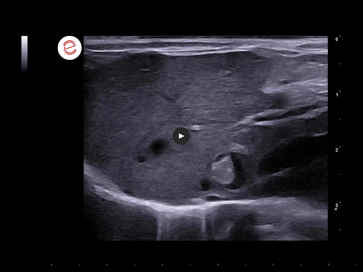
Transverse hepatic image from a right lateral coronal view.
At the junction of the central and left hepatic veins, a subacute thrombus with mixed echogenicity is present. This lesion causes critical stenosis and the development of acquired intrahepatic collateral vessels, visible as small transverse vascular structures.
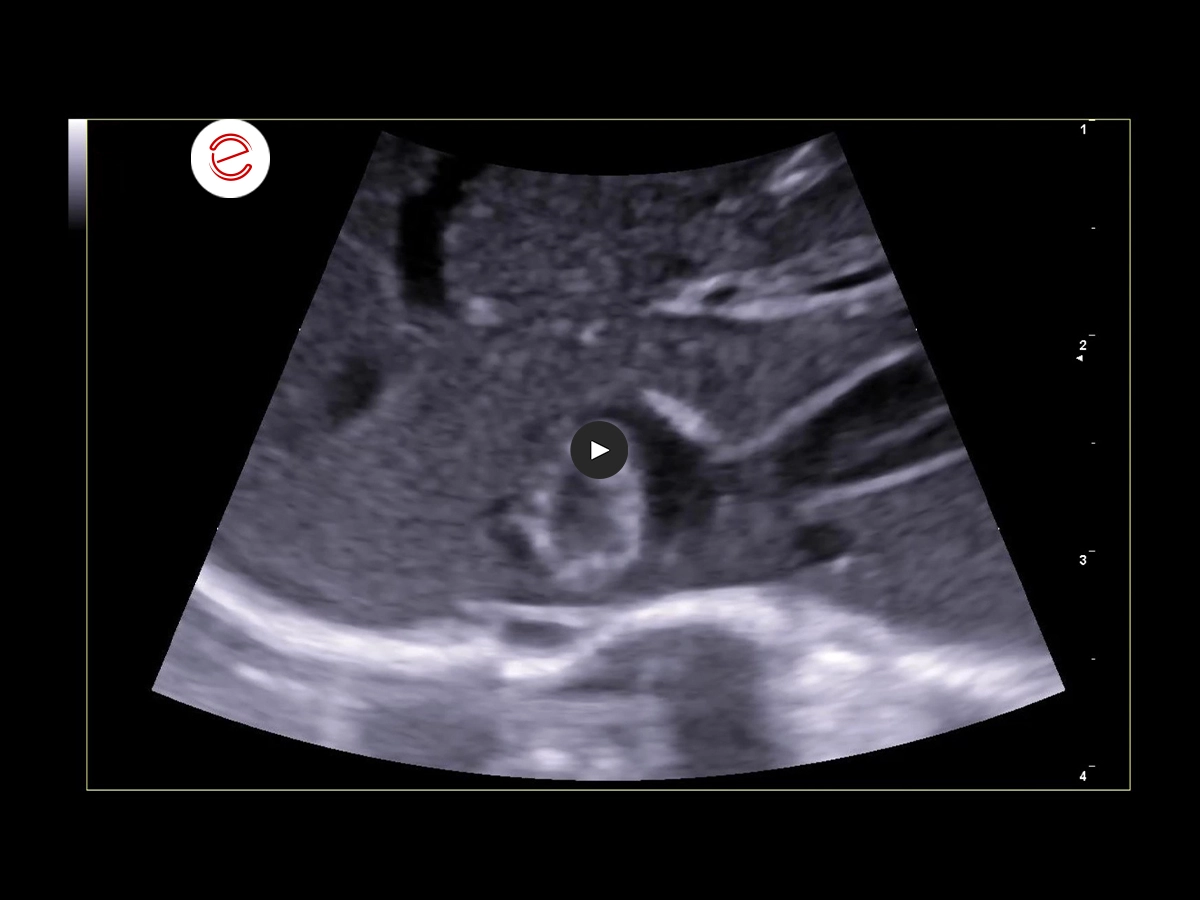
Transverse hepatic image with magnification of the hepatic vein junction.
Magnified view of the junction between the central and left hepatic veins, showing the subacute thrombus and collateral vessels. These findings are consistent with primary radicular Budd-Chiari syndrome and post-hepatic portal hypertension.
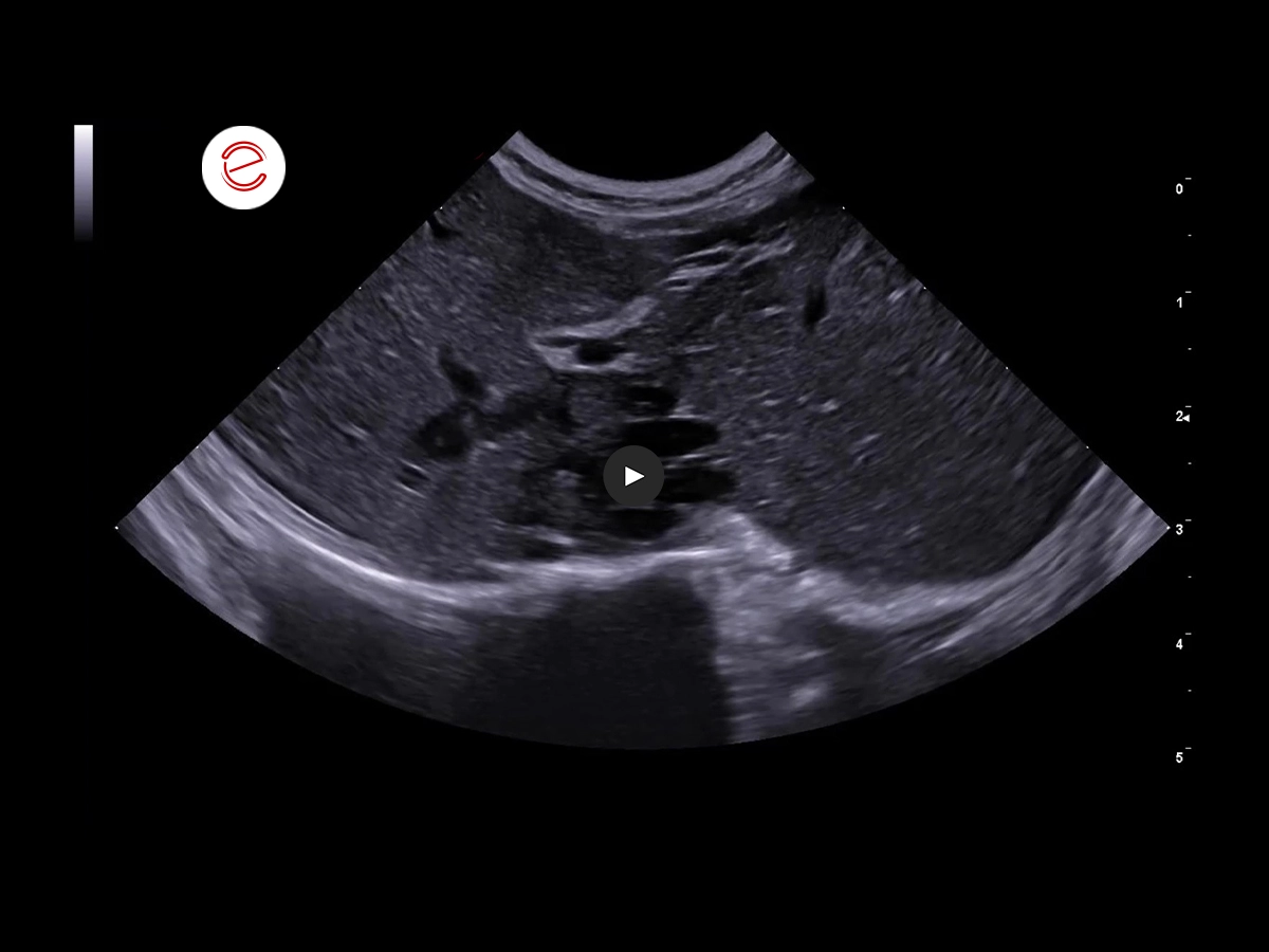
Transverse hepatic image from a right lateral coronal view.
After two months of antiplatelet therapy (clopidogrel), follow-up examination demonstrated a reduction in thrombus size, while the venous collateral circulation remained evident.
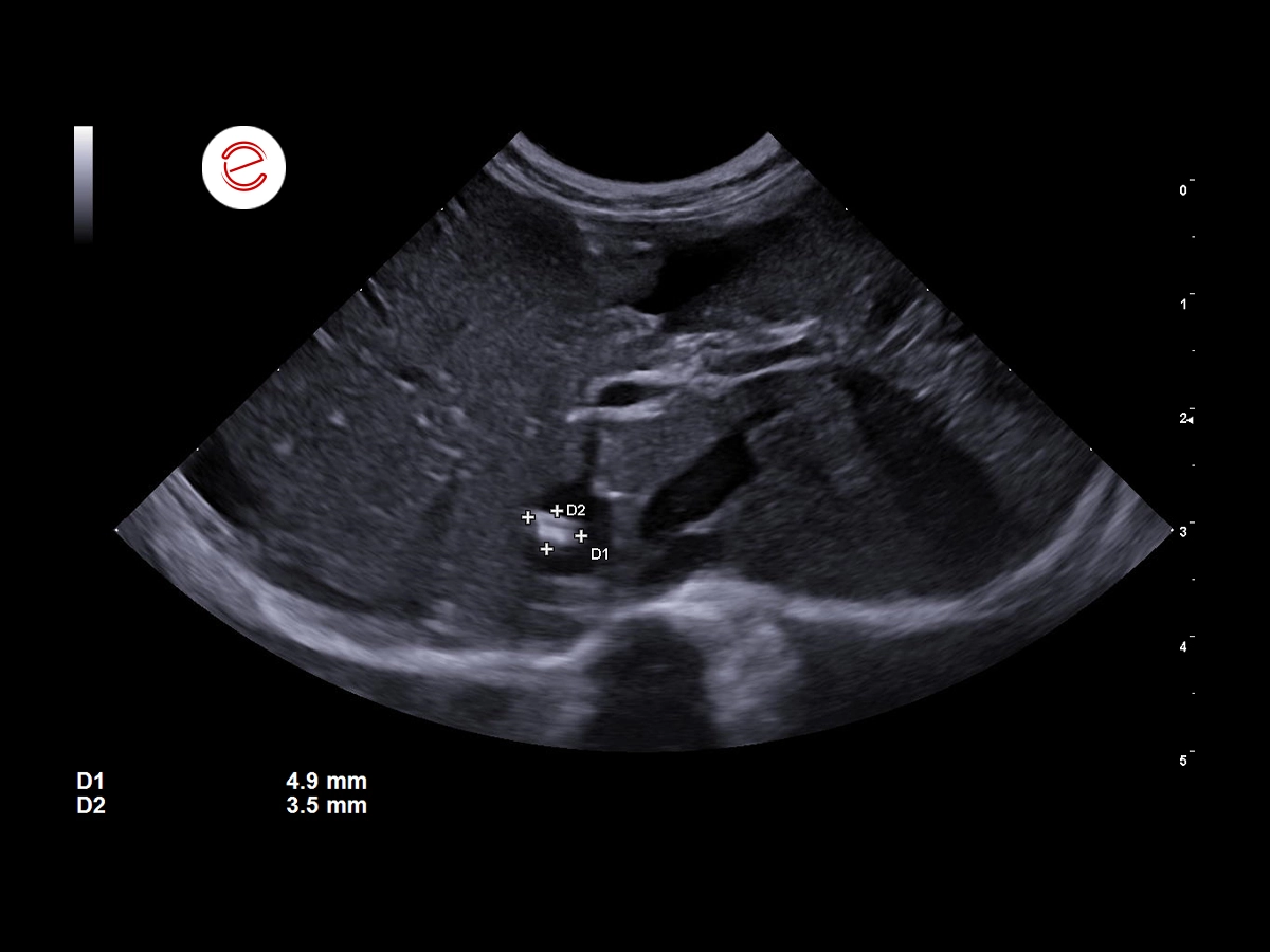
Transverse hepatic image from a right lateral coronal view.
The venous thrombus decreased in size and echogenicity and become adherent to the venous wall following treatment.
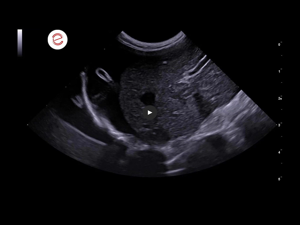
During the follow-up examination, a parasitic form of Dioctophyma renale was identified attached to the hepatic surface.
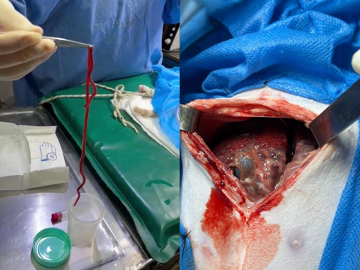
An exploratory laparotomy was performed to remove Dioctophyma renale. Surgical findings confirmed chronic peritoneal inflammation caused by parasite migration, which was considered one of the contributing factors to the abdominal effusion. Images courtesy of Dr. Juan Delgado Stagnares (Argentina).
Images were acquire with MyLab™Panther system.
Conclusions and Treatment
Brujo presented with a migratory form of Dioctophyma renale within the abdominal cavity, associated with systemic venous congestion impairing normal venous return to the thorax. Although the exact etiology of this condition remains unclear, previous reports have identified potential contributing factors, including congenital vascular webs affecting the caudal vena cava (Hehne et al.), veno-occlusive disease (Cave et al.), neoplastic conditions (Whitlock et al.), and congenital cardiac abnormalities. Knowledge of hepatic vascular disorders and thorough hemodynamic assessment are essential for the comprehensive evaluation of patients presenting with ascites, regardless of age.
Dr. Guadalupe Ranea, DVM, Postgraduate Professor at UBA and Co-Director of VetDoppler, Buenos Aires, Argentina.
MyLab is a trademark of Esaote spa.
Technology and features are device/configuration-dependent. Specifications subject to change without notice. Information might refer to products or modalities not yet approved in all countries. Product images are for illustrative purposes only. For further details, please contact your Esaote sales representative.
Other feline clinical cases you may be interested in
Explore the unique challenges encountered, the diagnostic procedures conducted, the solutions implemented, and the recommended treatments.

JULY 2026
Feline eosinophilic sclerosing fibroplasia
Gabriele Barella, DVM, PhD, MSc, “Centro Veterinario Doc4Pet Agno” veterinary center (CH)

MARCH 2026
Feline Pancreatitis Leading to Extrahepatic Biliary Obstruction and Jaundice
Dr Hélène Kolb, Associate Veterinarian / Medical Director of the Atlantia Veterinary Hospital, Member of the AFVAC Imaging Study Group (GEIM), Member…

JULY 2024
Neoplastic infiltrative process
Elisabetta Boz, DVM, Clinica Veterinaria Gran Sasso, Milan, Italy

