Adrenomegaly and bilateral nephropathy
Identification and history
- Name: Benjamin
- Report and medical history: canine, French bulldog, neutered male, 7 years old.
The patient showed slight dejection and required screening.
Diagnostics
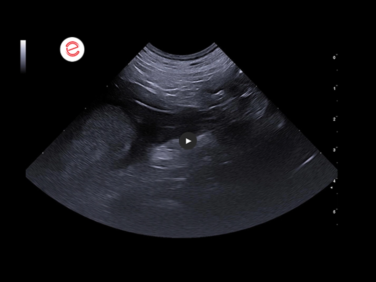
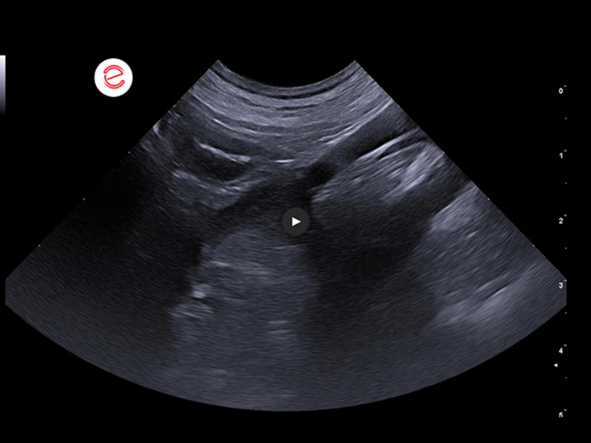
Inside the caudal vena cava in its pre-hepatic tract, an echogenic endoluminal structure was seen, globular in shape and parenchymatous in appearance, occupying 90% of the vascular lumen.
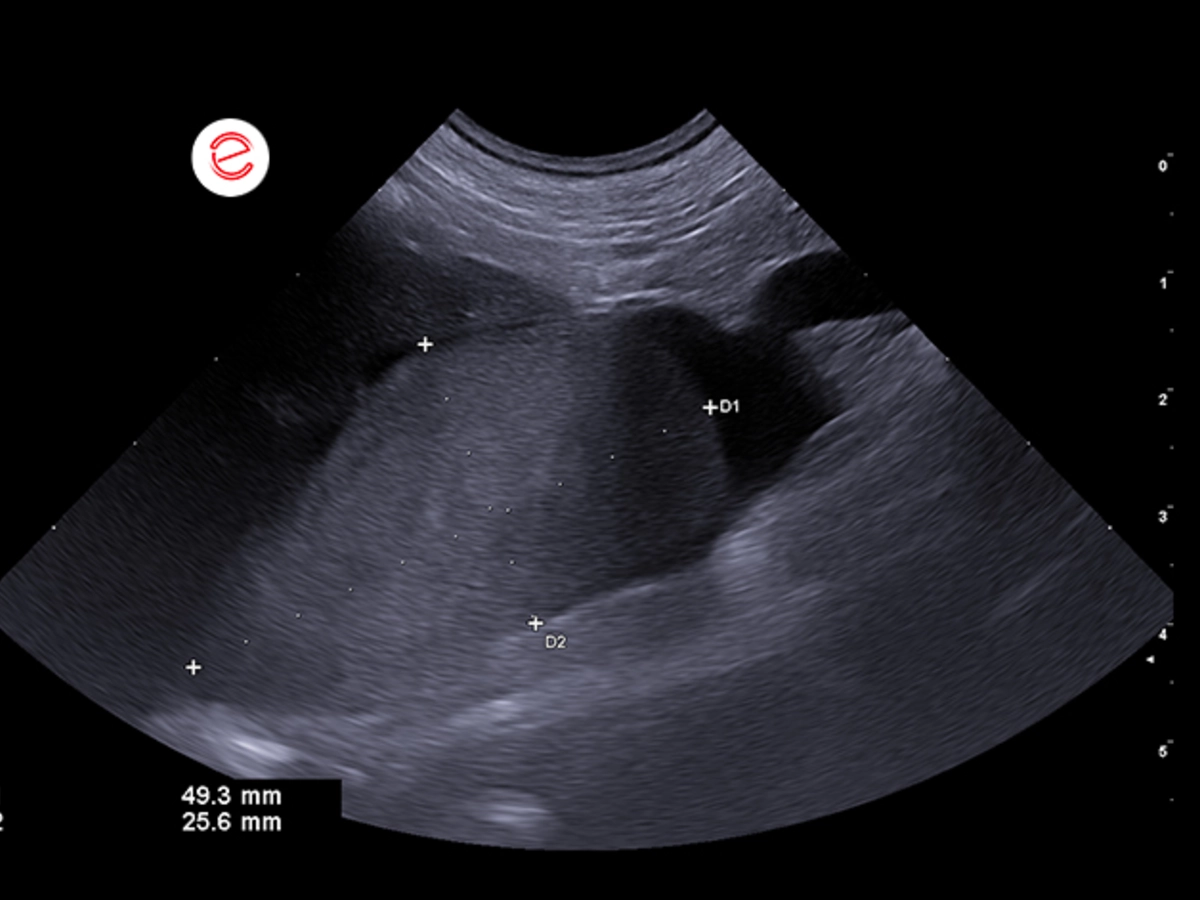
The maximum measurable dimensions of the mass were approximately 5 cm x 2.5 cm.
Kidneys within limits for size, regular profile.
Cortico-medullary distinction is slightly reduced due to a minor increase in bone marrow echogenicity.
Pelvis within normal limits.
Good distinction of the renal margins with respect to the surrounding tissues.
Peri-pelvic adipose tissue within normal limits.
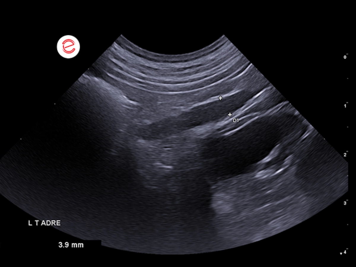
Left adrenal gland within the norm for size, regular shape and echostructure.
Thickness at the caudal pole of 3.9 mm.
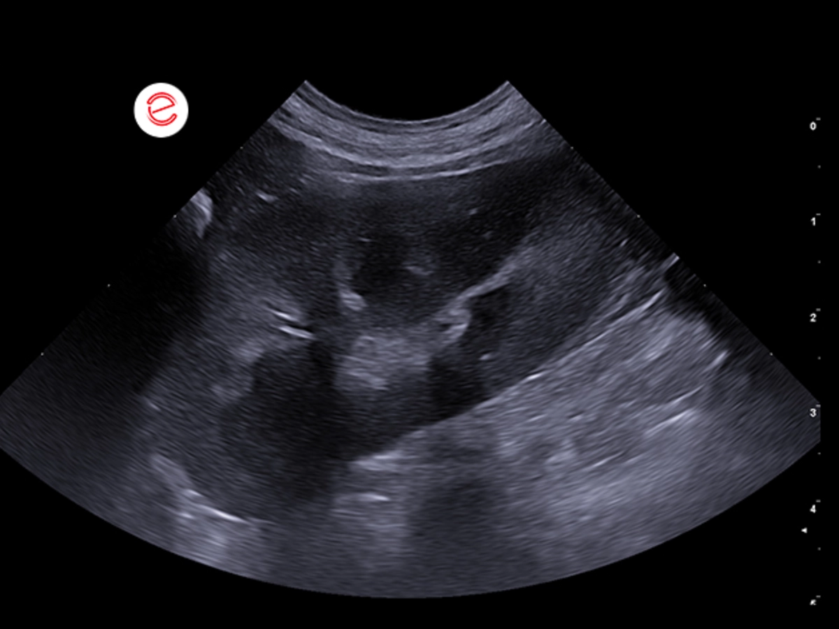
The right adrenal gland was increased in size with a globular appearance and poorly defined margins.
Echostructure slightly uneven.
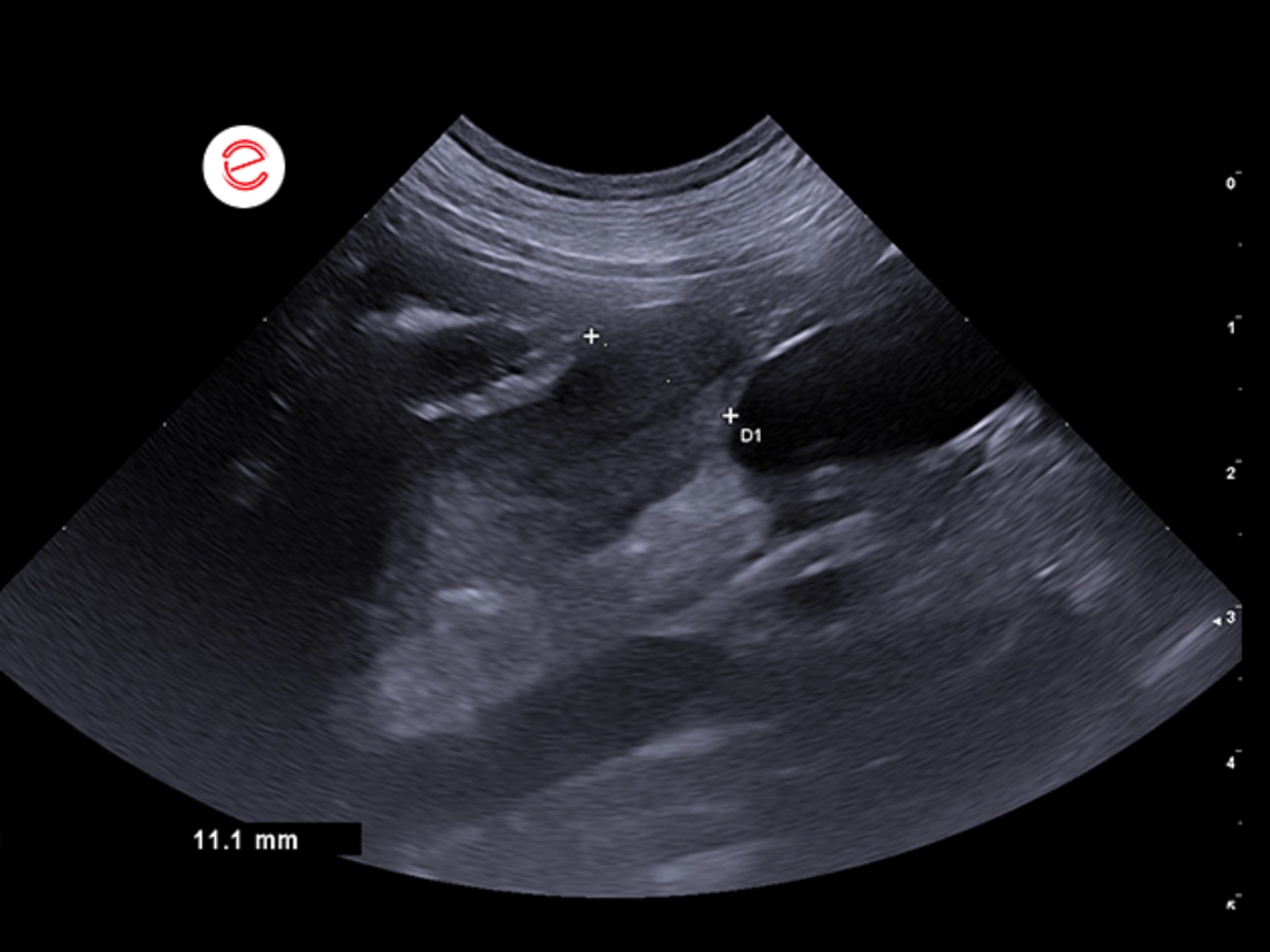
The thickness at the caudal pole was 11 mm.
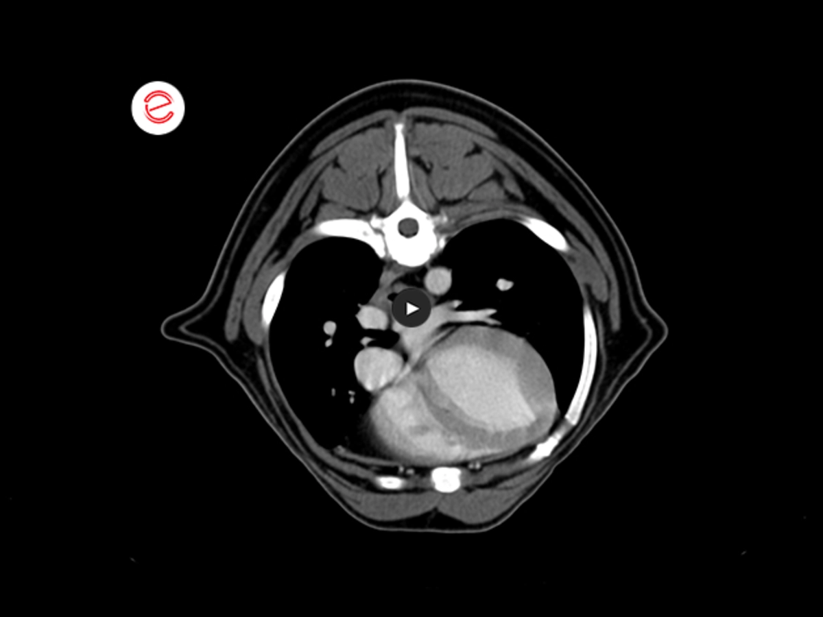
On CT examination, a right adrenal neoformation (1.2 cm maximum thickness in cross-section) was seen, globular in shape and characterized by marked and inhomogeneous enhancement.
This neoformation infiltrates the caudal vena cava, which was markedly increased in size with a focal filling defect in the post-contracted phase that extends for 9 cm and occupied almost the entire vascular lumen with initial caval collateral vessels along the periphery of the caudal vena cava.
A similar defect is associated in the right phrenic-abdominal vein.
Incidental presence of a small abnormal vessel that extends between the left gastric vein along the small curvature of the stomach to the diaphragm where it flexes medially entering the post-hepatic portion of the caudal vena cava through the phrenic vein.
Histological examination of the right adrenal gland and thrombus in vena cava was performed.
Macroscopy findings
A complete longitudinal section of the adrenal gland with the nodule and a cross-section of the vena cava with thrombus were examined.
Histology findings
The adrenal medulla was expanded and replaced by an unencapsulated, poorly demarcated, densely cellular, multilobulated neoplasm composed of polygonal cells arranged in nests and packages supported by a fine fibrovascular stroma, which compressed and/or infiltrated the surrounding cortical. The neoplastic cells had variably distinct margins, poorly moderated eosinophilic finely granular cytoplasm, a round, central/paracentral nucleus with finely dotted chromatin, and a distinct nucleolus. Moderate anisocytosis and anisokaryosis present. Mitoses on average fewer than 1 per 2.37 mm2. At the center of the neoplasm there were occasional areas of hemorrhage, fibrin and edema up to 0.5 mm in diameter and occasional macrophages filled with hemosiderin. In one of the sections, the infiltration of the wall of a large venous vessel (vena cava) with subtotal occlusion of the vascular lumen was evident.
Morphology findings
Adrenal gland, malignant pheochromocytoma with invasion and thrombosis of the vena cava.
Images were acquired using the MyLab™X90VET system.
Conclusions and treatment
- Right caudal vena cava thrombosis, coagulopathy/neoplastic thrombus.
- Right adrenomegaly: pheochromocytoma.
- Bilateral nephropathy with chronic aspects.
University Veterinary Hospital, Department of Diagnostic Imaging, University of Milan, Lodi.
MyLab is a trademark of Esaote spa.
Technology and features are device/configuration-dependent. Specifications subject to change without notice. Information might refer to products or modalities not yet approved in all countries. Product images are for illustrative purposes only. For further details, please contact your Esaote sales representative.
Other canine clinical cases you may be interested in
Discover the challenges faced, the examinations performed, the solutions adopted, and the treatments recommended.

MARCH 2025
Adrenal tumors
Carmelo Marco Bruno, DVM, specialist in pathology and clinic of companion animals, accredited with FSA and BOL
MAY 2024
Synovial Sarcoma
Daniel Sáez, DVM
Centro de Diagnóstico Veterinario Vetpoint®, Chile

MARCH 2024
Right atrial Hemangiosarcoma
Angeles Carrión DVM, Accre. AVEPA in Cardiology
Vetocardia, cardiología y ecografía veterinaria, Murcia, Spain
