Mammary intraductal carcinoma
Identification and history
- Name: Akira
- Report and medical history: canine, Siberian Husky, female neutered, 12 years old.
Approximately three months before presentation, a progressively enlarging subcutaneous mass developed in the right inguinal region, adjacent to the right inguinal mammary gland. Clinical examination revealed a firm, mobile, irregularly surfaced, and mildly painful subcutaneous lesion measuring ~6 cm. Bloodwork results were unremarkable.
Diagnostics
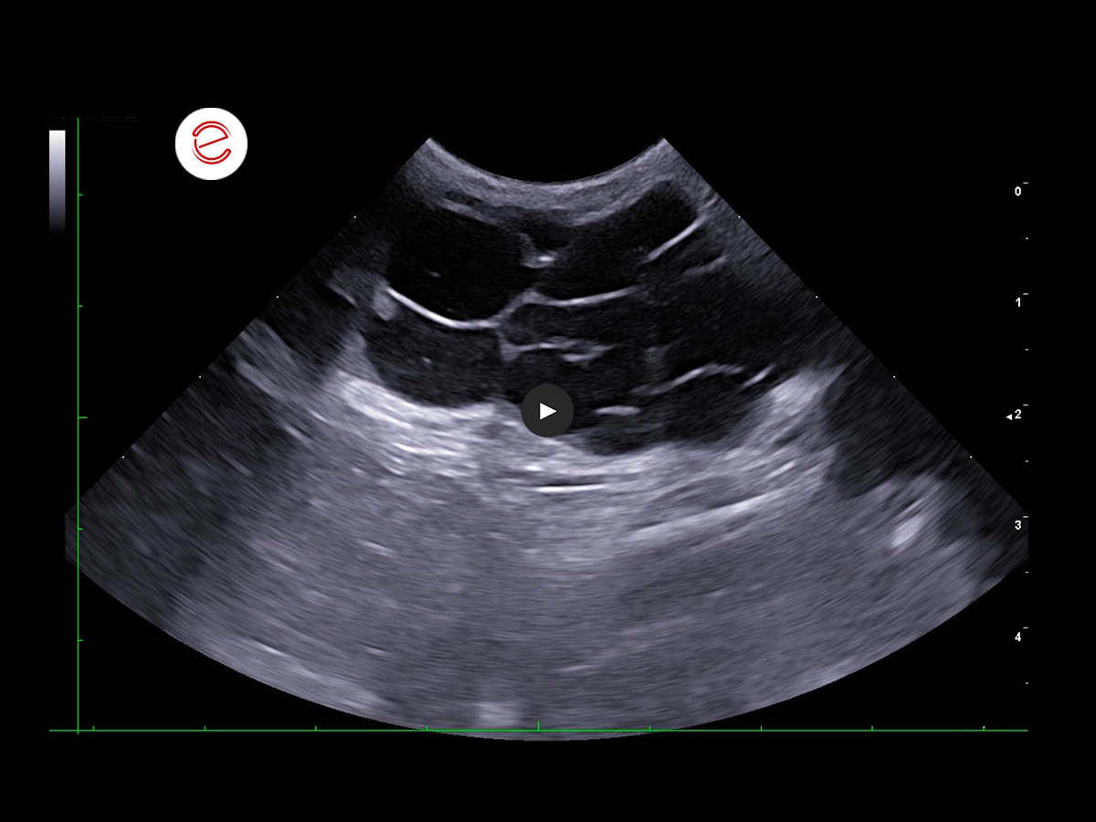
A multilobulated subcutaneous lesion consisting of multiple anechoic cavities with thin hyperechoic walls was observed in the right inguinal region, measuring up to 1.5 cm in diameter, and extending within the subcutaneous tissue in a cranio-caudal direction for approximately 7 cm.
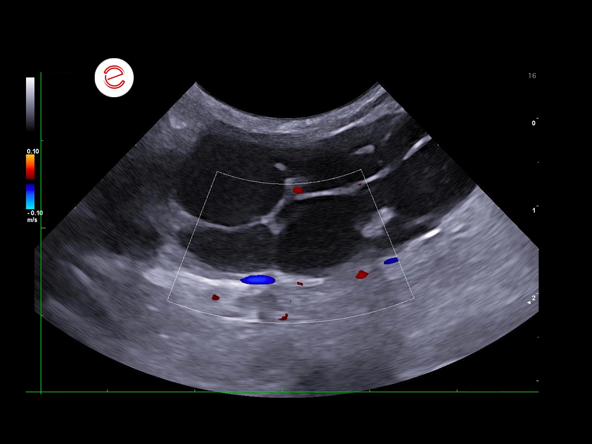
Doppler interrogation showed minimal peripheral vascularization.
Fine-needle aspiration yielded ~5 mL of clear, straw-colored fluid.
Images were acquired using the MyLab™X90VET system.
Cytological evaluation
Cytological evaluation revealed hemorrhagic samples with abundant macrophages containing hemosiderin and numerous erythrocytes, within a proteinaceous background. Findings were consistent with a cystic lesion with a hemorrhagic component.
After surgery, following an excisional biopsy of the lesion and regional lymph node, a histopathological examination revealed mammary intraductal carcinoma associated with ductal papillomatosis. The inguinal lymph node showed only mild follicular hyperplasia, and no evidence of metastatic disease.
Novel findings
This case provides a useful insight for clinicians encountering subcutaneous inguinal lesions with a multilobulated, cystic appearance on ultrasound. To the authors’ knowledge, this ultrasonographic presentation of mammary intraductal carcinoma has not been previously reported in dogs. Awareness of this unusual manifestation may help guide differential diagnoses and emphasize the need to consider neoplasms in inguinal subcutaneous lesions that appear cystic.
Chiara Donà, DVM, PhD, Faculty of Veterinary Medicine, University of Milan, Italy

MyLab is a trademark of Esaote spa.
Technology and features are device/configuration-dependent. Specifications subject to change without notice. Information might refer to products or modalities not yet approved in all countries. Product images are for illustrative purposes only. For further details, please contact your Esaote sales representative.
Other canine clinical cases you may be interested in
Discover the challenges faced, the examinations performed, the solutions adopted, and the treatments recommended.

NOVEMBER 2025
Myxomatous mitral valve disease and heartworm infection
Paul A. Cardenio DVM, MS, DPCCP, DPCVS-CA, Medical Director, PetLandia Veterinary Clinic, City of Malolos, Bulacan, Philippines

JULY 2025
Fibroliposarcoma and malignant melanoma
Reynaldo Buan Jr., DVM, MSc - Buan Veterinary Practice Pampanga, Filippine
MAY 2025
Adrenomegaly and bilateral nephropathy
University Veterinary Hospital, Department of Diagnostic Imaging, University of Milan, Lodi.
