Adrenal adenoma
Identification and history
- Name: Nina
- Report and medical history: Dog, Short-haired Dachshund, Female, 10 years old
Ultrasound checkup required for weight loss, PU/PD, recurrent vomiting, especially after ingestion of extra-diet foods
Diagnostics
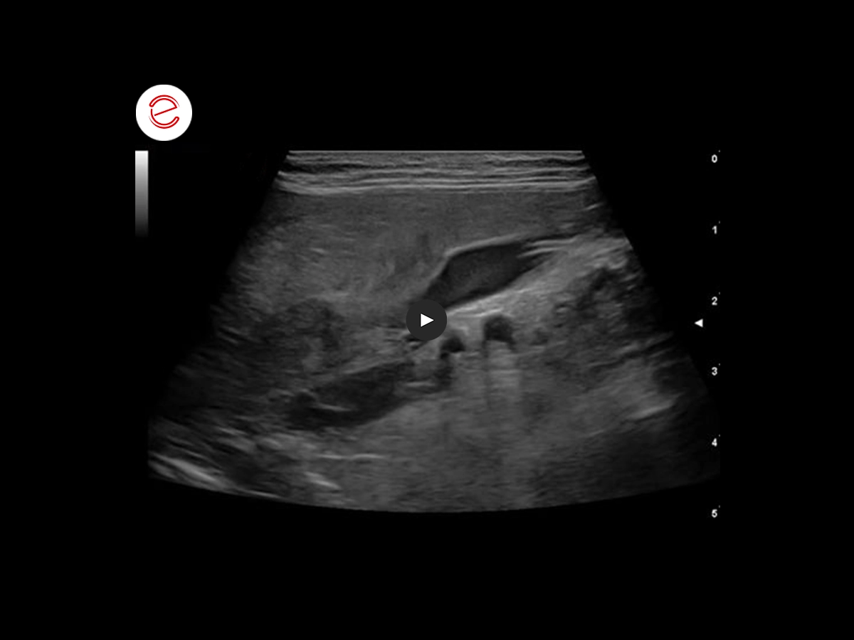
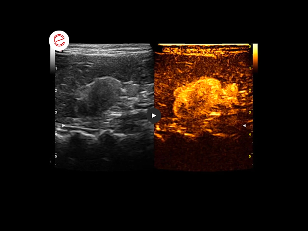
Neoformation in the right adrenal gland with inhomogeneous echo structure, associated with evident other echogenic neoformation within the right phrenic/abdominal artery and caudal vena cava in the prehepatic tract.
In the video we can observe, caudally to the celiac and cranial mesenteric arteries, the contralateral adrenal gland with tendentially decreased volume.
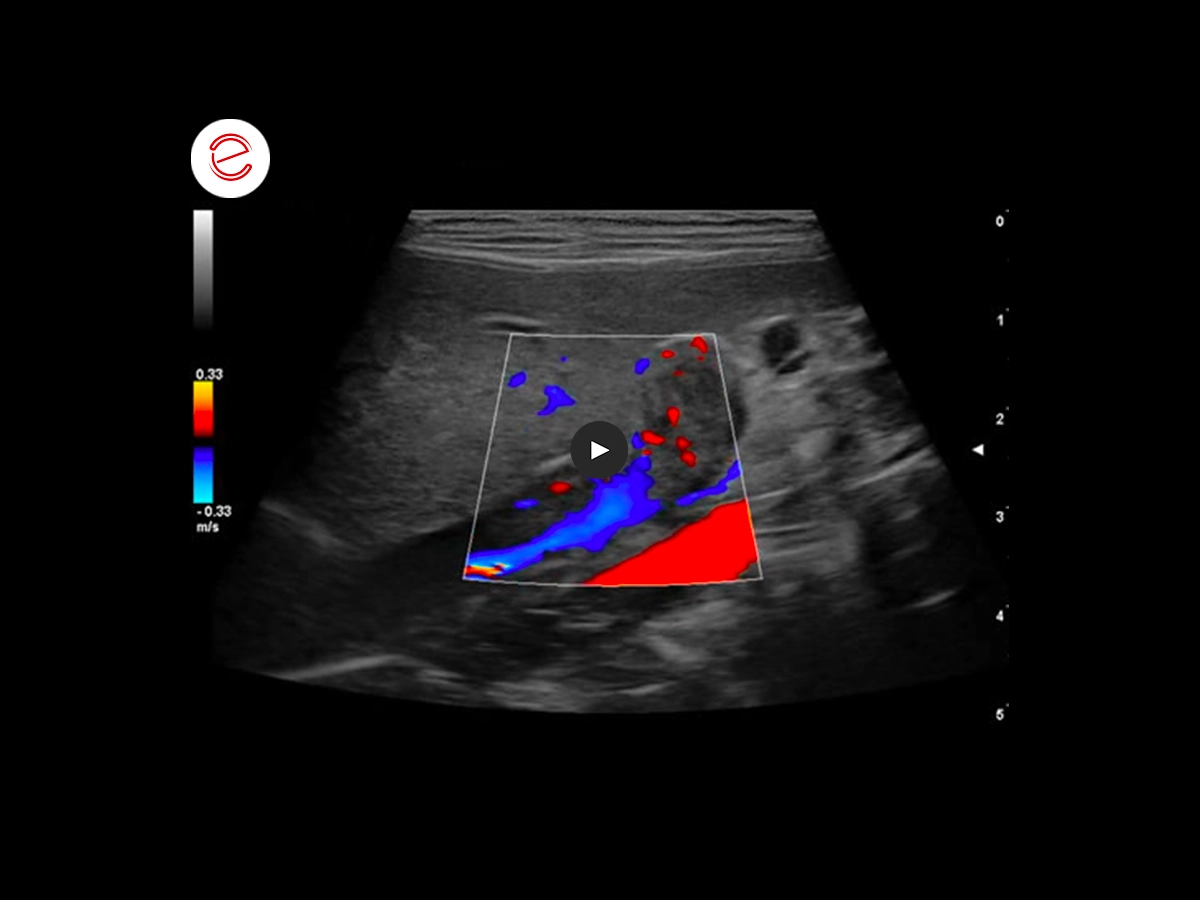
Study of the vascularization of the mass. We notice both arterial and venous flows within the adrenal neoformation are present. We can further make out a turbulence in the caudal vena cava cranially to the formation linked to the increase in the post-stenotic flow velocity.
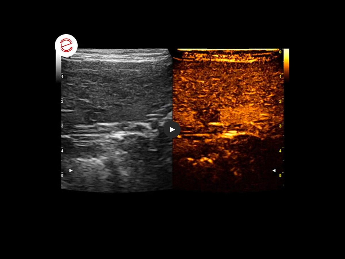
Image composed using the CEUS technique performed with contrast medium (sulfur hexafluoride microbubbles) at a dose of 0.04 ml/kg through the cephalic vein, showing poor acquisition probably due to a thrombotic neovascularization.
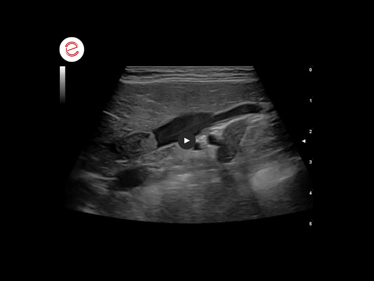
A suspect hepatic hyperechoic lesion has been studied which, according to the CEUS method, shows a wash-in and wash-out phase at the same time as the remaining hepatic vascularization, not compatible with metastatic lesion.
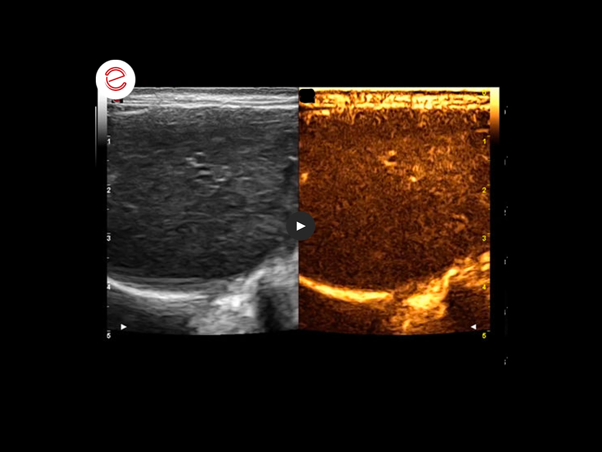
The right adrenal gland shows progressive and homogeneous wash-in with centripetal pattern. We can notice a slow and orderly discharge of the contrast medium in the subsequent phase. The echo contrast graphic image can be referenced in the first hypothesis to adrenal neoplasia with secondary venous thrombosis. Blood chemistry analyses, urine tests, and low and then high dose suppression tests with dexamethasone confirmed independent ACTH hypercortisolism.
Conclusions and treatment
Adrenal cortisol-secreting adenoma/adenocarcinoma.
Adrenalectomy surgery to be considered.
Sergio Fanfoni, DVM, Clinica Veterinaria Santa Cristina, Monte San Savino, Italy, SCIVAC Coordinator.
MyLab is a trademark of Esaote spa.
Technology and features are device/configuration-dependent. Specifications subject to change without notice. Information might refer to products or modalities not yet approved in all countries. Product images are for illustrative purposes only. For further details, please contact your Esaote sales representative.
Other canine clinical cases you may be interested in
Discover the challenges faced, the examinations performed, the solutions adopted, and the treatments recommended.

JANUARY 2026
Mammary intraductal carcinoma
Chiara Donà, DVM, PhD, Faculty of Veterinary Medicine, University of Milan, Italy

NOVEMBER 2025
Myxomatous mitral valve disease and heartworm infection
Paul A. Cardenio DVM, MS, DPCCP, DPCVS-CA, Medical Director, PetLandia Veterinary Clinic, City of Malolos, Bulacan, Philippines

JULY 2025
Fibroliposarcoma and malignant melanoma
Reynaldo Buan Jr., DVM, MSc - Buan Veterinary Practice Pampanga, Filippine
