Monocytic ehrlichiosis
Identification and history
- Name: Suzy
- Report and medical history: canine, Shih Tzu, male, 10 years old.
Suzy, a 10-year-old male Shih Tzu dog, presented with phthisis bulbi and complete blindness in the right eye. The left eye had normal vision. Three days later, the owner noticed that the left eye appeared red and glazed. The dog had been bumping into objects for a few days. Hyphema and anterior uveitis were found in the left eye. A blood test revealed anemia with thrombocytopenia. Acute blindness in the left eye was diagnosed based on a negative menace response, dazzle reflex, and pupillary light reflex. A diagnosis can be made relatively easily based on a physical and ophthalmological examination. It is especially important to perform a detailed examination of the fundus using an ophthalmoscope. In this case, other tests were necessary, such as B‑mode ocular ultrasound with a hockey probe and high‑frequency transducer (7.5–25 MHz), performed with the transcorneal technique in the horizontal plane. In addition to ocular disease, this dog was concurrently diagnosed with canine monocytic ehrlichiosis.
Diagnostics
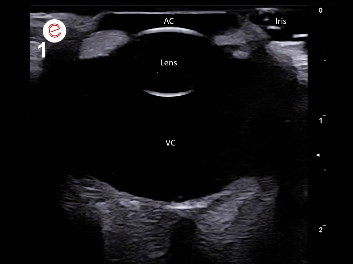
Figure 1. An ultrasonography image of a normal ocular globe showing that the anterior chamber (AC), and the lens and vitreous chamber (VC) appear anechoic. The iris shows a hyperechoic band-like structure.
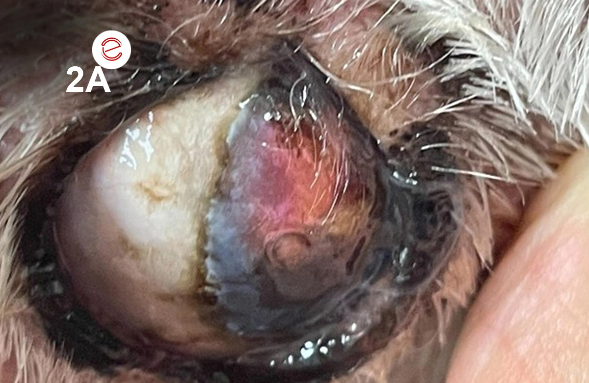
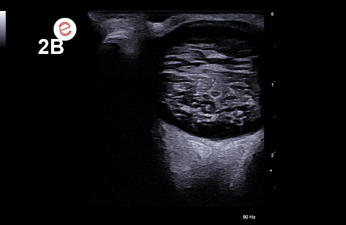
Figure 2A: Phthisis bulbi and complete blindness in the right eye.
Figure 2B: The ultrasonography image shows ocular abnormalities and shrunken globe with extensive calcification and loss of the normal eye shape.
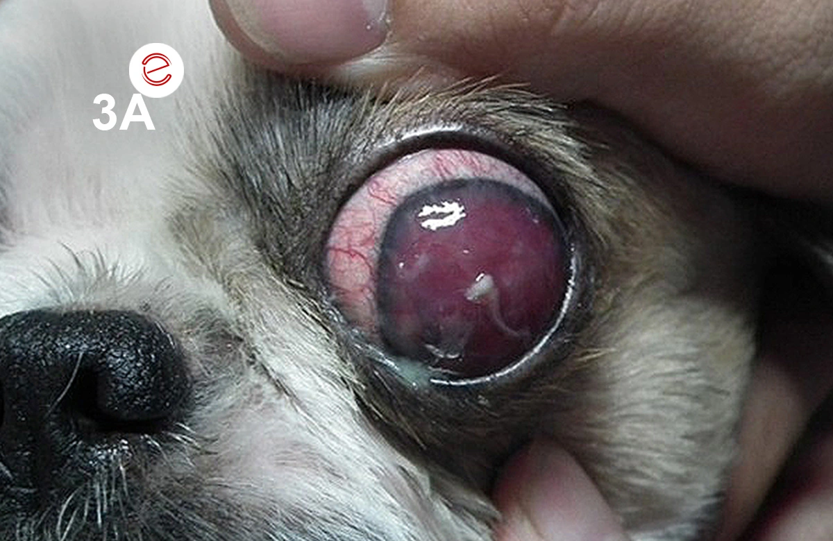
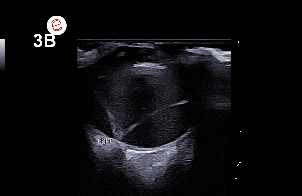
Figure 3A: Hyphema and anterior uveitis were found in the left eye.
Figure 3B: The ultrasonography shows a thin, hyperechoic, curvilinear structure in the vitreous chamber, resembling a V-shaped echo. There is subretinal hemorrhage, which is seen as a homogenous hypoechoic area.
Images were acquired with MyLab™FOX.
Treatment
This case was treated with topical atropine and topical prednisolone acetate to control the uveitis. Doxycycline at 5 mg/kg PO every 12 hours, administered daily for 4 consecutive weeks, was the systemic treatment of choice for ehrlichiosis. Medical therapy is usually preferred, especially for exudative retinal detachments. Treatment is directed at resolving the underlying disease; corticosteroids at immunosuppressive doses are generally administered, typically prednisolone at 1–2 mg/kg every 12–24 hours for 1–2 months. Hyphema and anterior uveitis with complete retinal detachment, causing acute blindness, were suspected to be induced by canine monocytic ehrlichiosis.
Discussion
In this case, the fundus could not be properly examined; therefore, ocular ultrasonography is the method of choice for evaluating the position fi the retina (Esteban Martín, 2007). For diagnosing retinal detachment using ultrasound, B‑mode and a high‑frequency transducer are primarily employed. Total detachment appears as a continuous, thin, curvilinear, hyperechoic structure in the posterior chamber that can be traced to the optic nerve head, often showing a V- or Y- shaped configuration, as showing in Fig 3 (Cho, 2021; Grahn et al., 2007).
Conclusions
Examinatining the fundus using an ophthalmoscope is especially important, as this can reveal out‑of‑focus areas of the retina. Ocular ultrasonography is the method of choice for identifying intraocular lesions when ocular opacity precludes direct visualization. Ophthalmoscopic examination with a high‑frequency hockey probe (7.5–25 MHz) is well suited for ocular ultrasound examination.
Arnon Chumcomlue, Ophthalmology Clinic, Arnon Pet Hospital, Chiang Rai

References
Cho, J. (2021). Ocular ultrasound abnormalities and optic nerve sheath diameter in dogs and cats. Veterinary Clinics of North America: Small Animal Practice, 51(6). doi.org/10.1016/j.cvsm.2021.07.010
Esteban Martín, J. (2007). Atlas de oftalmología clínica del perro y del gato (1st ed.). Servet.
Grahn, B. H., Barnes, L. D., Breaux, C. B., & Sandmeyer, L. S. (2007). Chronic retinal detachment and giant retinal tears in 34 dogs: Outcome comparison of no treatment, topical medical therapy, and retinal reattachment after vitrectomy. Canadian Veterinary Journal, 48(10).
MyLab is a trademark of Esaote spa.
Technology and features are device/configuration-dependent. Specifications subject to change without notice. Information might refer to products or modalities not yet approved in all countries. Product images are for illustrative purposes only. For further details, please contact your Esaote sales representative.
Other canine clinical cases you may be interested in
Discover the challenges faced, the examinations performed, the solutions adopted, and the treatments recommended.

JANUARY 2026
Mammary intraductal carcinoma
Chiara Donà, DVM, PhD, Faculty of Veterinary Medicine, University of Milan, Italy

NOVEMBER 2025
Myxomatous mitral valve disease and heartworm infection
Paul A. Cardenio DVM, MS, DPCCP, DPCVS-CA, Medical Director, PetLandia Veterinary Clinic, City of Malolos, Bulacan, Philippines

JULY 2025
Fibroliposarcoma and malignant melanoma
Reynaldo Buan Jr., DVM, MSc - Buan Veterinary Practice Pampanga, Filippine
