Adrenal tumors
Identification and history
- Name: Jackie
- Report and medical history: dog, spayed female, crossbreed, 10 years old, 19 kg.
The patient has been losing weight for some time with increased abdominal volume.
Cardiology consultation was requested for suspected ascites.
Diagnostics
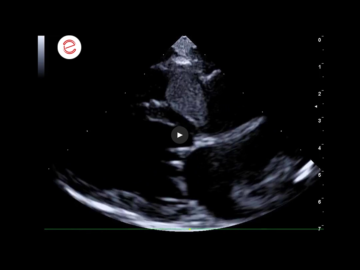
Right parasternal long-axis four-chamber view, Standard 1.
Normal atrial and ventricular volume.
Nodular neoplasm in the right atrium protruding into the right ventricle.
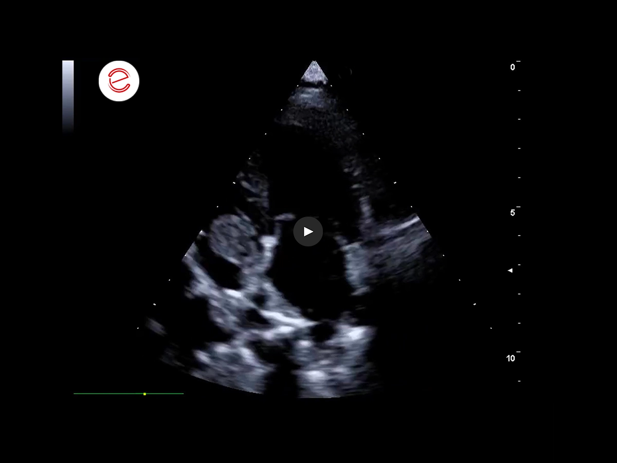
Right parasternal short-axis view, bicaval projection.
Nodular neoplasm in the right atrium which appears to originate from the caudal vena cava.
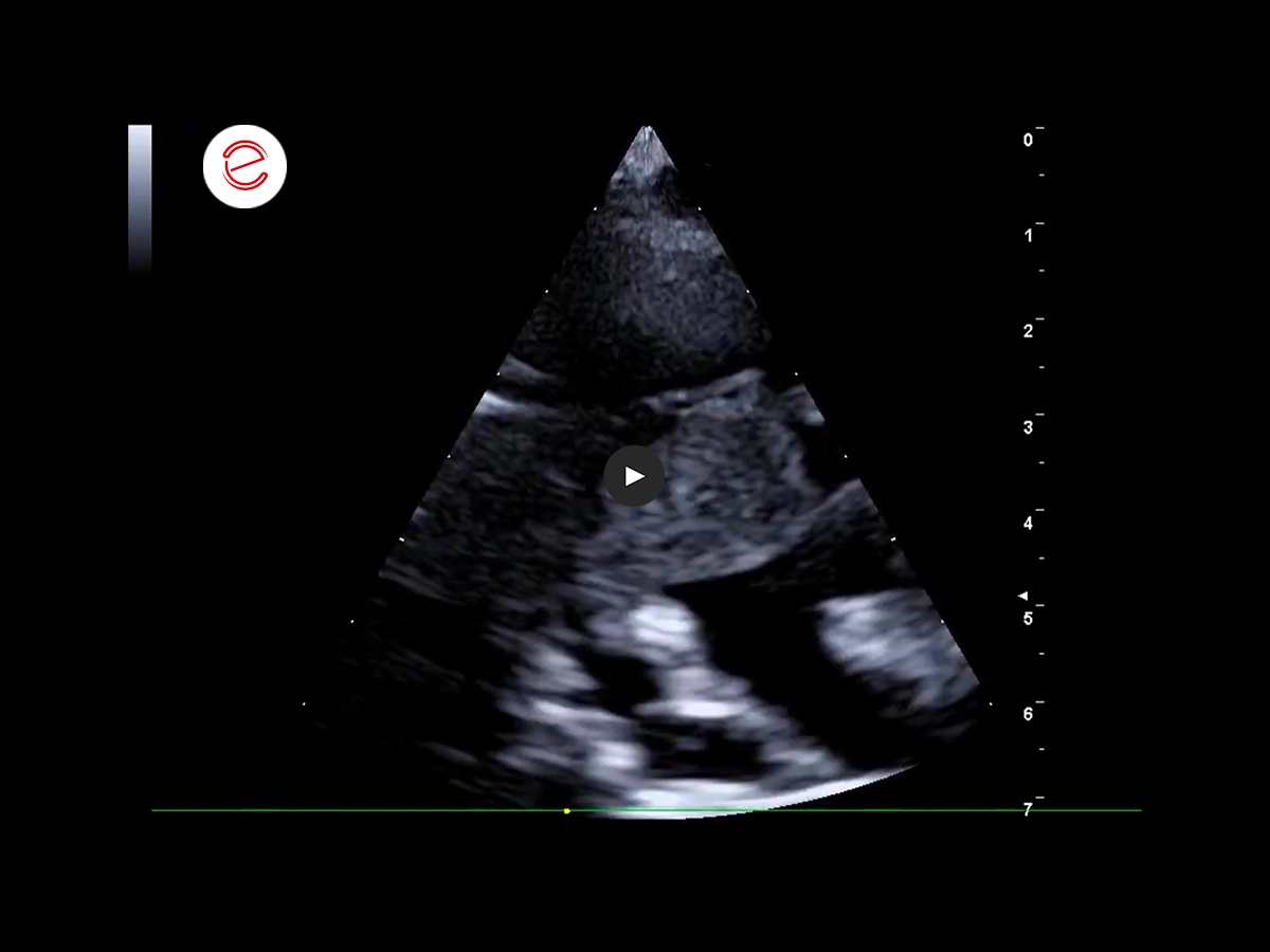
Left parasternal apical four-chamber view.
Normal atrial and ventricular volume.
Nodular neoplasm in the right atrium protruding into the right ventricle.
Scan of the right adrenal gland projection area with the patient in the left lateral decubitus position.
Neoplasm of approx. 3 cm invading the phrenico-abdominal vein.
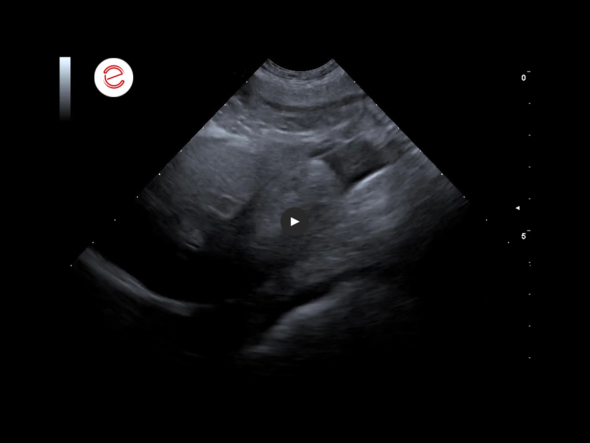
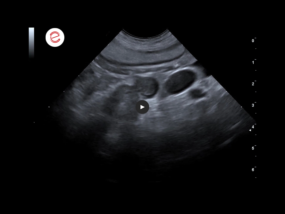
Hepato-caval scan with the patient in the left lateral decubitus position.
Intraluminal neoplasm occluding the vena cava with vascular ectasia.
Rounded liver lobes and presence of abdominal effusion.
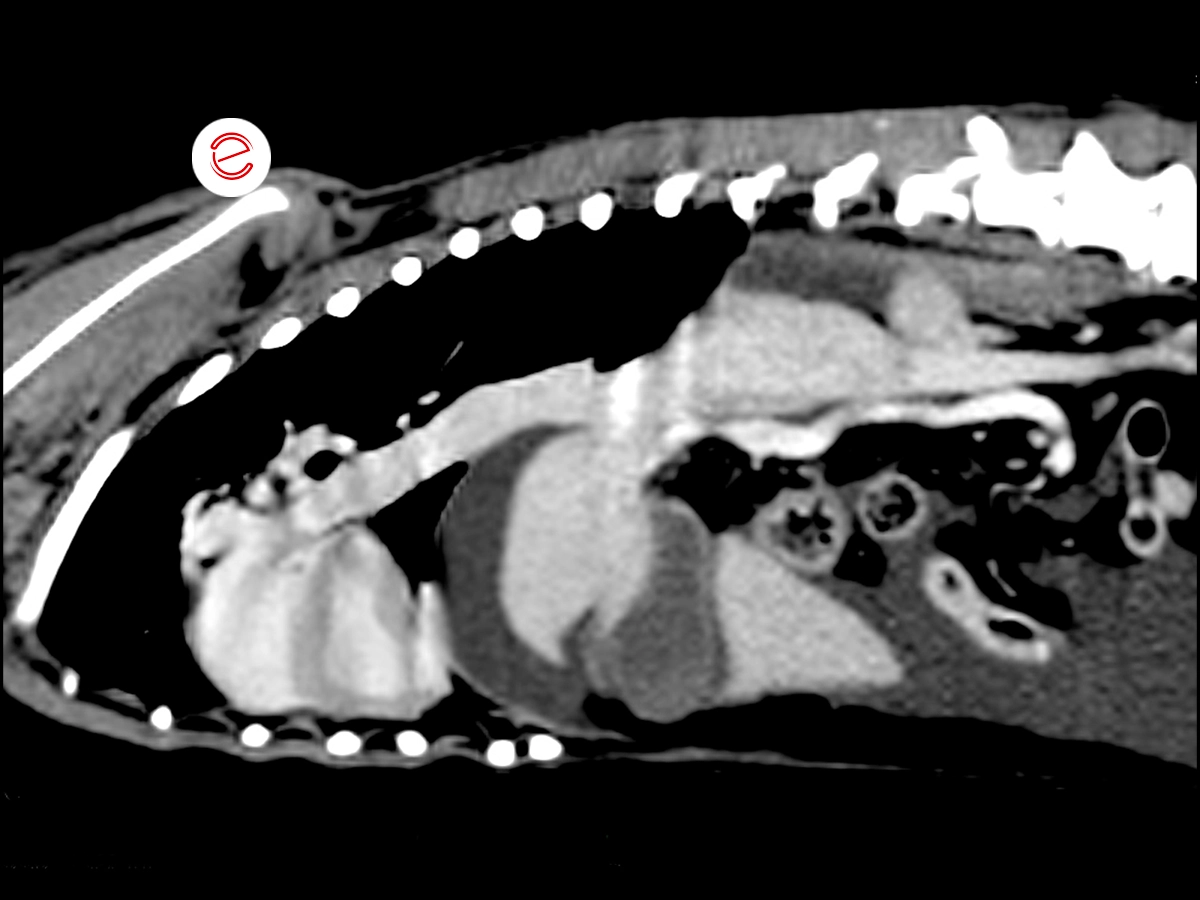
Sagittal-plane CT scan with contrast agent.
Neoplasm of the right adrenal gland with ill-defined margins and heterogeneous appearance, characterized by mild/moderate and heterogeneous contrast agent uptake.
An intraluminal extension was observed at the level of the phrenico-abdominal and ipsilateral cranial abdominal veins and caudal vena cava, extending cranially in the right atrium (approx. 29x26x164 mm).
Images were acquired using the MyLab™Omega eXP VET system.
Conclusions and treatment
Adrenal tumors can invade the caudal vena cava through the phrenico-abdominal vein; this phenomenon has been observed in approximately 11% of carcinomas and 50% of pheochromocytomas. In extreme cases, the intravascular growth may reach the right heart and cause blockage of vena cava venous return (Budd-Chiari like syndrome).
Surgical intervention may be a viable option due to the inability of the tumor to adhere to the vessel walls, which would allow the surgeon to remove the neoplasm through an abdominal approach.
Carmelo Marco Bruno, DVM, specialist in pathology and clinic of companion animals, accredited with FSA and BOL

MyLab is a trademark of Esaote spa.
Technology and features are device/configuration-dependent. Specifications subject to change without notice. Information might refer to products or modalities not yet approved in all countries. Product images are for illustrative purposes only. For further details, please contact your Esaote sales representative.
Other canine clinical cases you may be interested in
Discover the challenges faced, the examinations performed, the solutions adopted, and the treatments recommended.

JANUARY 2026
Mammary intraductal carcinoma
Chiara Donà, DVM, PhD, Faculty of Veterinary Medicine, University of Milan, Italy

NOVEMBER 2025
Myxomatous mitral valve disease and heartworm infection
Paul A. Cardenio DVM, MS, DPCCP, DPCVS-CA, Medical Director, PetLandia Veterinary Clinic, City of Malolos, Bulacan, Philippines

JULY 2025
Fibroliposarcoma and malignant melanoma
Reynaldo Buan Jr., DVM, MSc - Buan Veterinary Practice Pampanga, Filippine
