
 Asia Pacific (English)




Feline eosinophilic sclerosing fibroplasia
Identification and history
- Name: Kilo
- Report and medical history: European long-haired cat, netred male, 2 years old.
Clinical examination for chronic vomiting, anorexia, and weight loss.
Diagnostics
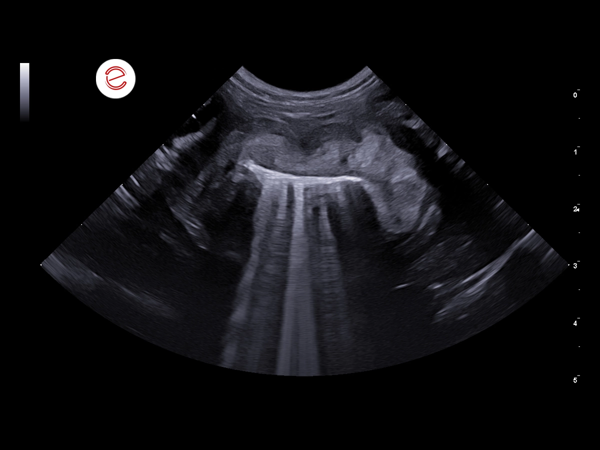
Right transverse retrocostal scan.
Ascending duodenum: severe parietal thickening is evident, with an overall inhomogeneous, hyperechogenic wall. The layering is partially preserved (severe alterations of the inner layers corresponding to the mucosa and submucosa, while the outer portion of the tunica muscularis layer is partially preserved).
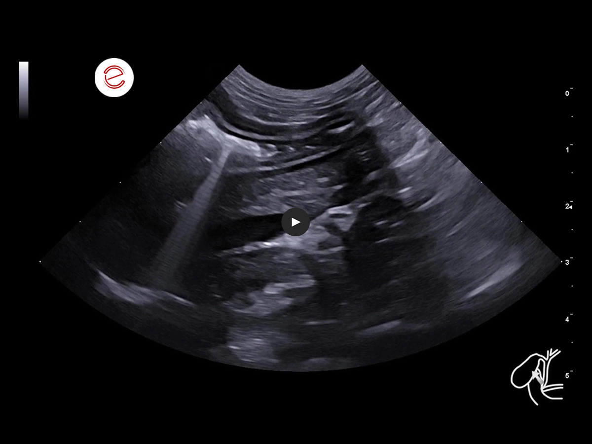

Right longitudinal retrocostal scan.
The ascending duodenum is slightly distended due to minimal fluid and gas content: severe asymmetric transmural parietal thickening is evident. The layering is partially preserved (severe alterations of the inner mucosal and submucolas layers, while the outer portion of the tunica muscularis is partially preserved). This thickening continues with the ultrasound showing a normal-looking descending segment of duodenum.
Fine-needle aspiration (FNA) of the duodenal wall was performed: inconclusive result.
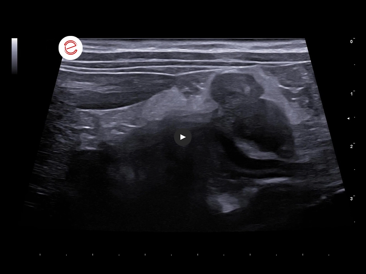
Longitudinal right retrocostal scan.
The gastric lymph node is moderately enlarged, with irregular, well-defined borders and an inhomogeneous hypoechogenic structure. Th hilum is not visible. Moderate reactivity of the adjacent peritoneal adipose tissue. Vascular Doppler signal was not assessed.
Fine-needle aspiration (FNA) of the gastric lymph node was performed
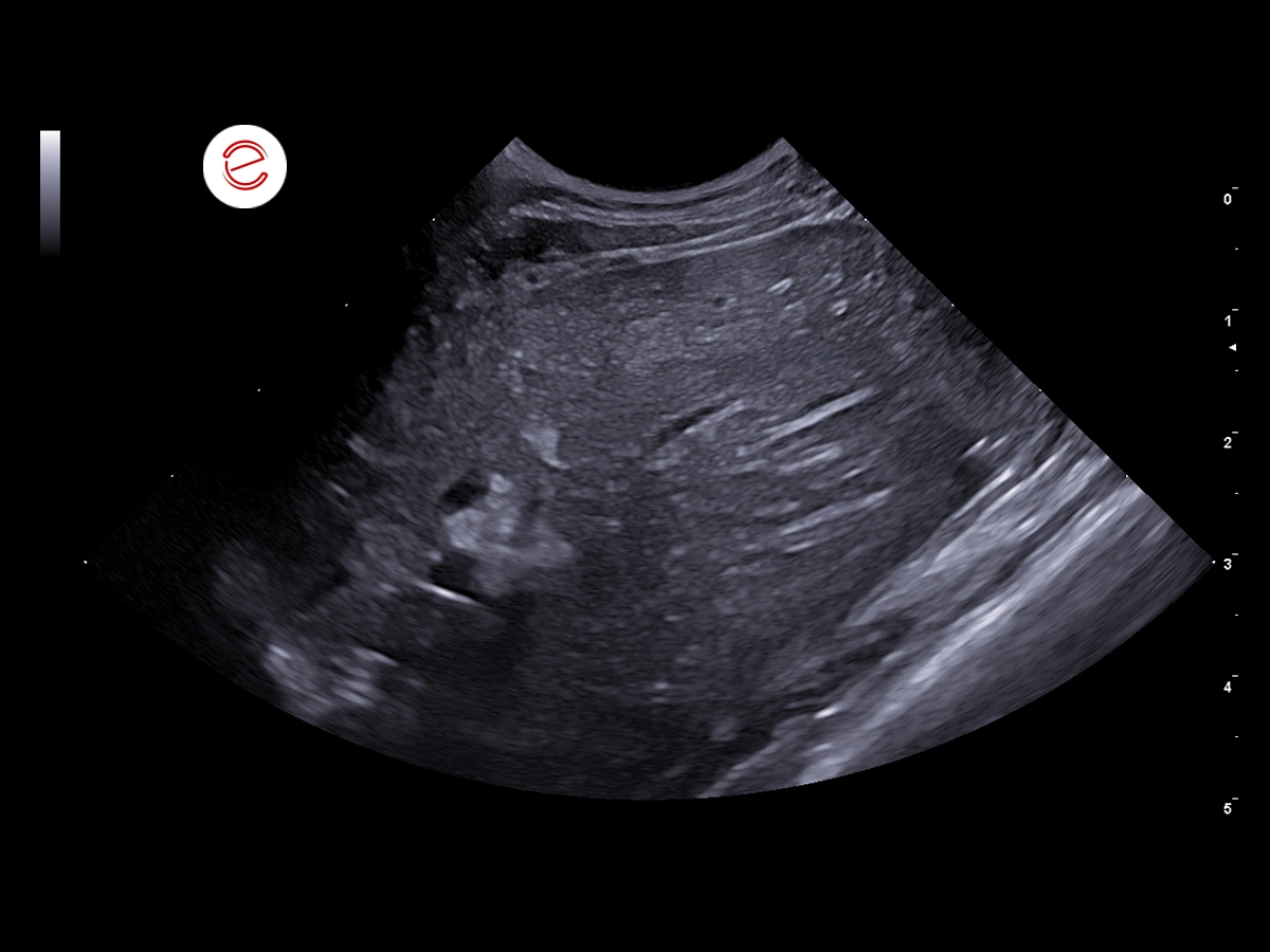
Left transverse intercostal scan.
The liver shows normal echogenicity and homogeneous appearance. Normal vascular pattern.
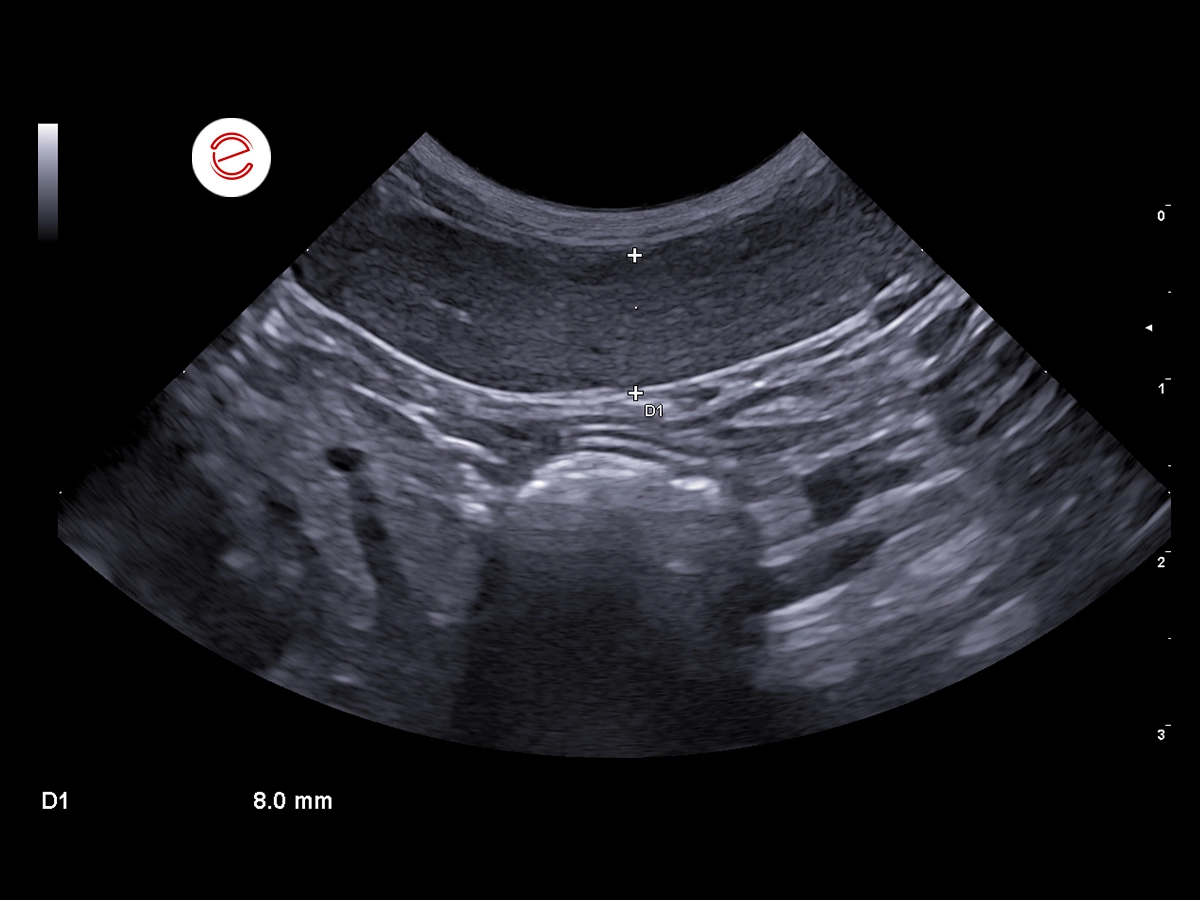
Left longitudinal retrocostal scan.
The spleen is normal in size, with regular margins and a homogeneous hyperechogenic echotexture.
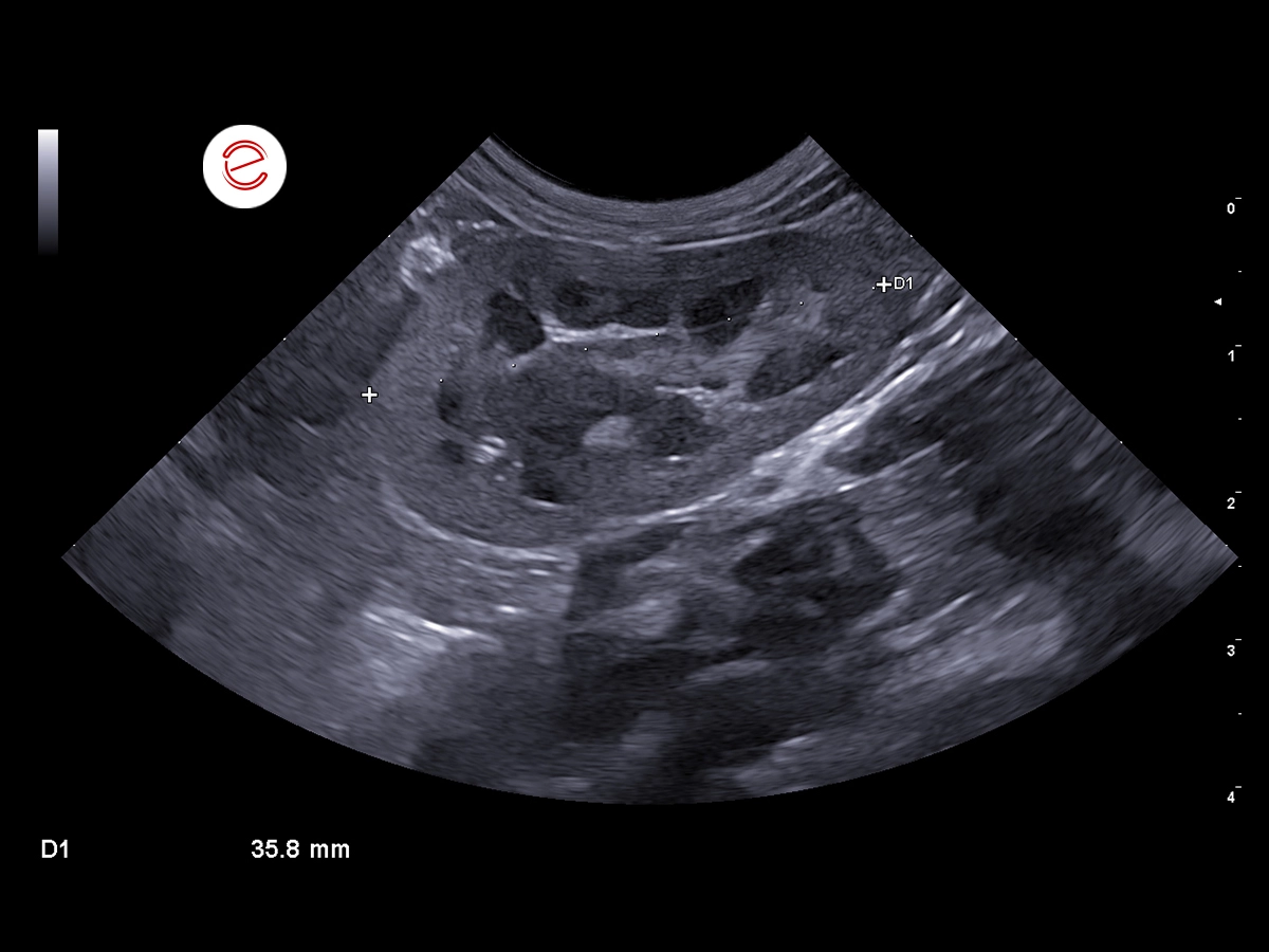
Left longitudinal scan.
The left kidney is normal in size with regular borders and a homogeneous, mildly hyperechogenic cortex. Corticomedullary definition is preserved.
Images were acquired with MyLab™Panther
Conclusions and Treatment
- The parietal thickening of the ascending duodenum shows no signs of occlusion, and is therefore consistent with a neoplastic process (DD: round cell neoplasm, less likely carcinoma, granulomatous enteritis, or feline eosinophilic sclerosing fibroplasia)
- A full-thickness biopsy sample of the duodenal wall was performed
- Result: feline eosinophilic sclerosing fibroplasia
Histological sections revealed numerous eosinophilic granulocytes infiltrating the lamina propria, alongside smaller numbers of plasma cells, lymphocytes, and neutrophilic granulocytes. The lining epithelium is intact, but there is focal infiltration by rare lymphocytes. Numerous proliferating fibroblasts embedded in abundant collagen and rare neutrophilic granulocytes were observed in the submucosa.
Gabriele Barella, DVM, PhD, MSc, “Centro Veterinario Doc4Pet Agno” veterinary center (CH)

MyLab is a trademark of Esaote spa.
Technology and features are device/configuration-dependent. Specifications subject to change without notice. Information might refer to products or modalities not yet approved in all countries. Product images are for illustrative purposes only. For further details, please contact your Esaote sales representative.
Other feline clinical cases you may be interested in
Explore the unique challenges encountered, the diagnostic procedures conducted, the solutions implemented, and the recommended treatments.

SEPTEMBER 2026
Budd-Chiari Syndrome and Dioctophyma renale
Dr. Guadalupe Ranea, DVM, Postgraduate Professor at UBA and Co-Director of VetDoppler, Buenos Aires, Argentina.

MARCH 2026
Feline Pancreatitis Leading to Extrahepatic Biliary Obstruction and Jaundice
Dr Hélène Kolb, Associate Veterinarian / Medical Director of the Atlantia Veterinary Hospital, Member of the AFVAC Imaging Study Group (GEIM), Member…

JULY 2024
Neoplastic infiltrative process
Elisabetta Boz, DVM, Clinica Veterinaria Gran Sasso, Milan, Italy

