Chronic fibrotic hepatopathy and myocardial granuloma
Identification and history
- Name: Mueller
- Report and medical history: Spix Macaw (Cyanopsitta spixii), male, 11 years old.
The bird is part of a conservation project and each bird within the facility is checked every year. This patient had a history of a chronic liver disease and was rechecked two months later due to respiratory problems.
A blood test showed a normal WBC count with heterophilia. Biochemistry showed increased level ofUric Acid, Calcium, cholesterol, AST, LDH, Total Protein, and a severe increase in Triglyceride levels. The troponin level was within the normal range.
Cloacal microbiology detected escherichia coli, but no fungi or yeast were cultured.
Diagnostics
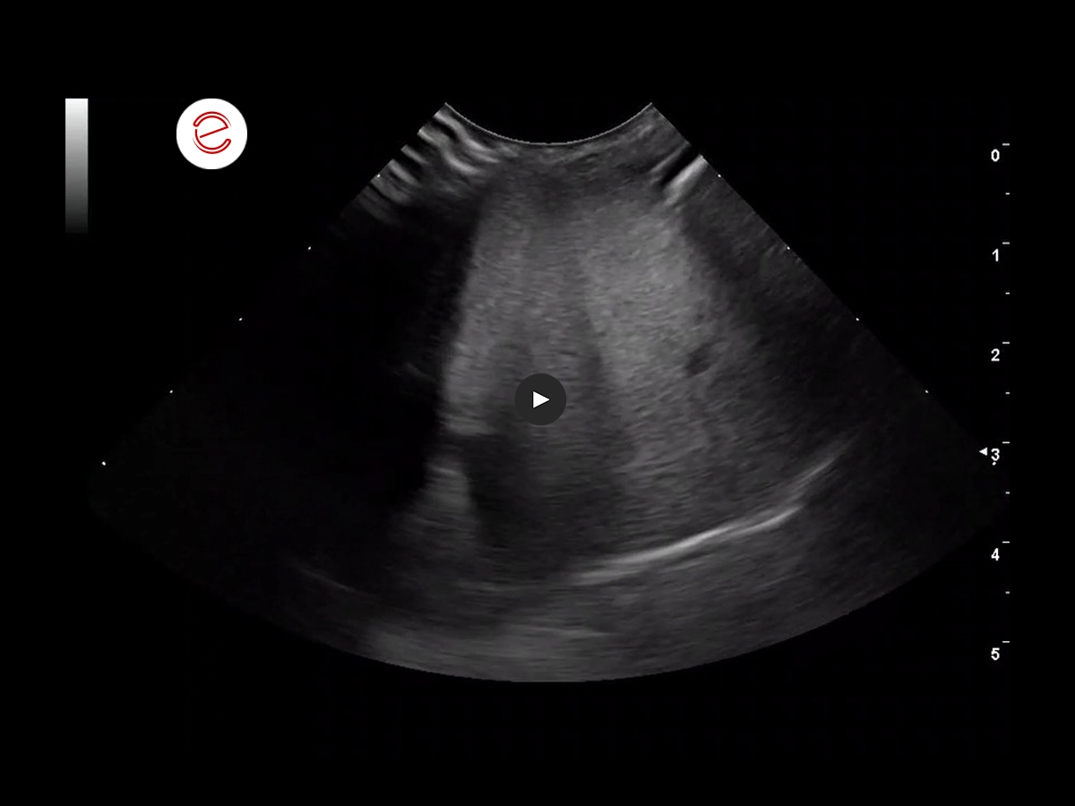
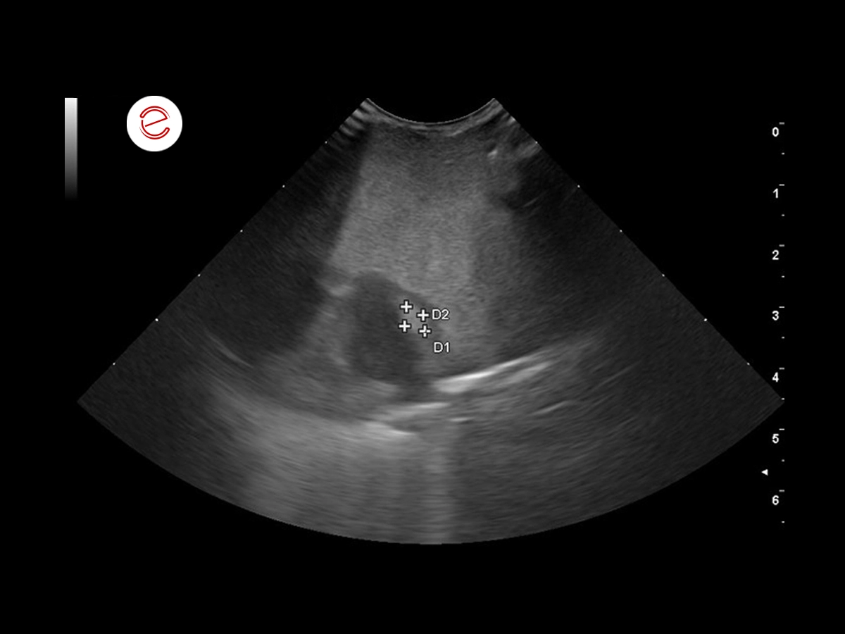
2 months before the respiratory issues: the larger right liver lobe is visible in the sagittal ventromedial view. The liver appears dense and heterogeneous, and the small hepatic vessels appear compressed.
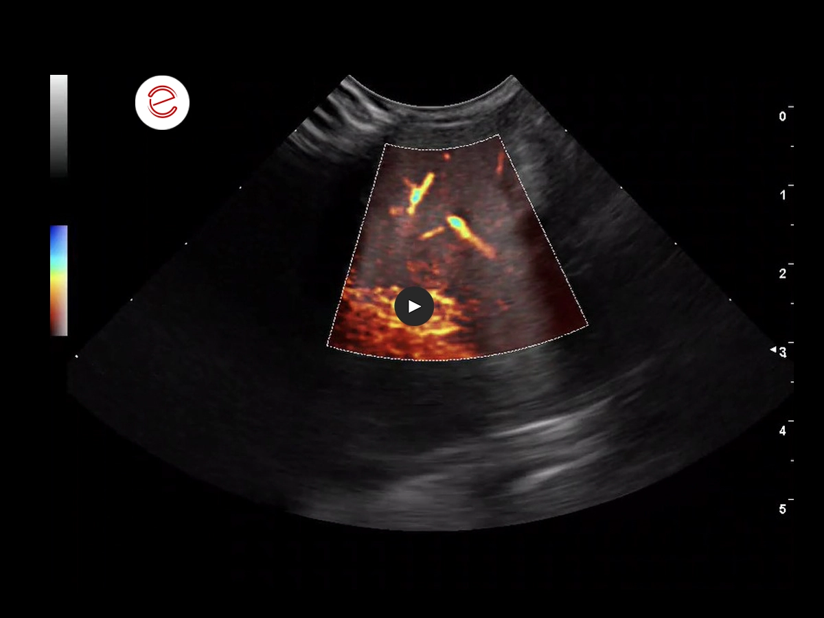
The microV function outlines the compressed vessels.
Histopathology from an endoscopy-guided liver biopsy revealed liver fibrosis.
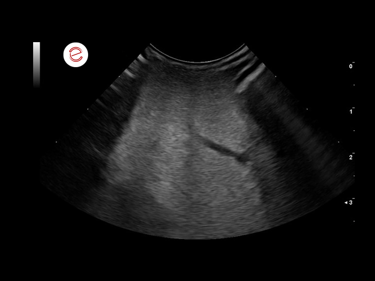
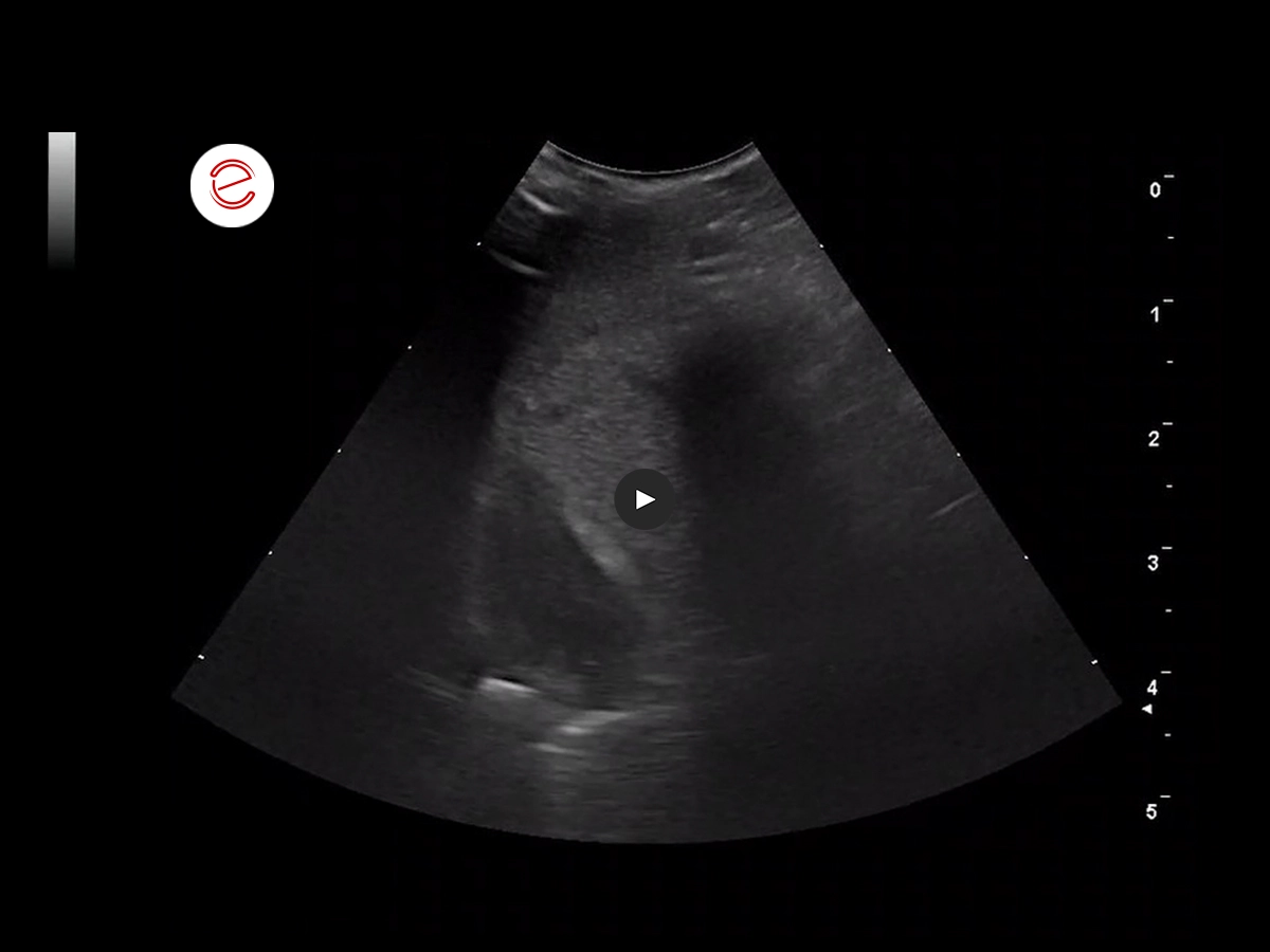
2 months later, the bird was presented for a follow up, mainly due to the sudden onset of respiratory problems.
There was a mild improvement of the liver.
Treatment: LIV52 (an ayurvedic remedy well established in avian medicine), Silymarin, Ursodeoxycholic Acid, dietary change.

In birds, during the celomic examination it is essential to examine the heart as well.
The absence of a diaphragm in birds and this peculiar anatomical situation ensures that the heart is in direct contact with the liver. Infections in birds travel not only in the conventional hematogenous way, but also directly from organ to organ.
In this case, a hyperechoic oval structure was visible in the lateral wall of the left ventricle.
Measurement of the mass within the myocardium of the left ventricle.
Differential diagnoses include a granuloma (fungal or bacterial), neoplasia and hematoma.
After 2 months of treatment, the bird showed improvement clinically and in the ultrasound examination the mass/granuloma has visibly reduced in size.
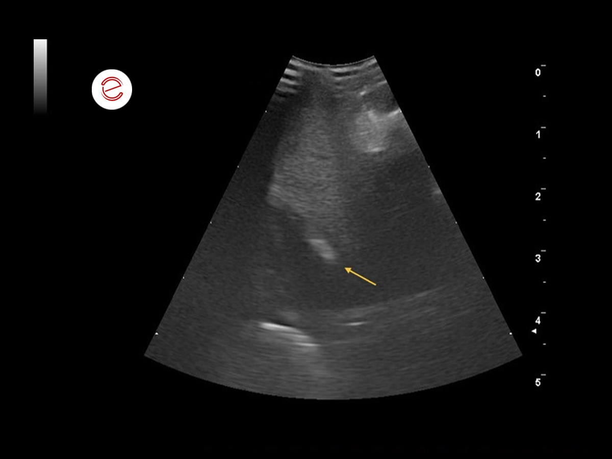
The yellow arrow shows the scar tissue that is now present.
Images were acquired with MyLab™Omega eXP VET
Conclusions and treatment
The granuloma in the myocardium was an incidental finding. The respiratory disorders resolved within the first few weeks of treatment with antibiotics, hepatoprotection and heart medication. The bird was rechecked monthly until the granuloma resolved after two months, although scar tissue was still visible within the muscle. The suspected cause of the granuloma was a systemic Escherichia coli infection resulting in bacteremia.
Five months later, the bird is behaving normally with no sign of disease. It is still on heart medication and a different diet, as well as receiving liver protection.
Petra Schnitzer, med vet, GPCert-ExAP, MANZCVS, Dipl ACEPM, Master Small Animal Ultrasound, DIVAS, University of Milan, Freelancer

MyLab is a trademark of Esaote spa.
Technology and features are device/configuration-dependent. Specifications subject to change without notice. Information might refer to products or modalities not yet approved in all countries. Product images are for illustrative purposes only. For further details, please contact your Esaote sales representative.
Other exotics clinical cases you may be interested in
Discover the challenges faced, the examinations performed, the solutions adopted, and the treatments recommended.