Synovial Sarcoma
Identification and history
- Name: Brownie
- Report and medical history: Dog, Mongrel, 8 ys, Male neutered.
The patient had chronic progressive lameness of the left hind limb with mild, non-painful periarticular swelling and mild pain on extension and flexion. Drawer and tibial compression tests were inconclusive. The rest of the physical examination was within normal limits.
The presumptive diagnosis was a cranial cruciate ligament rupture.
Diagnostics
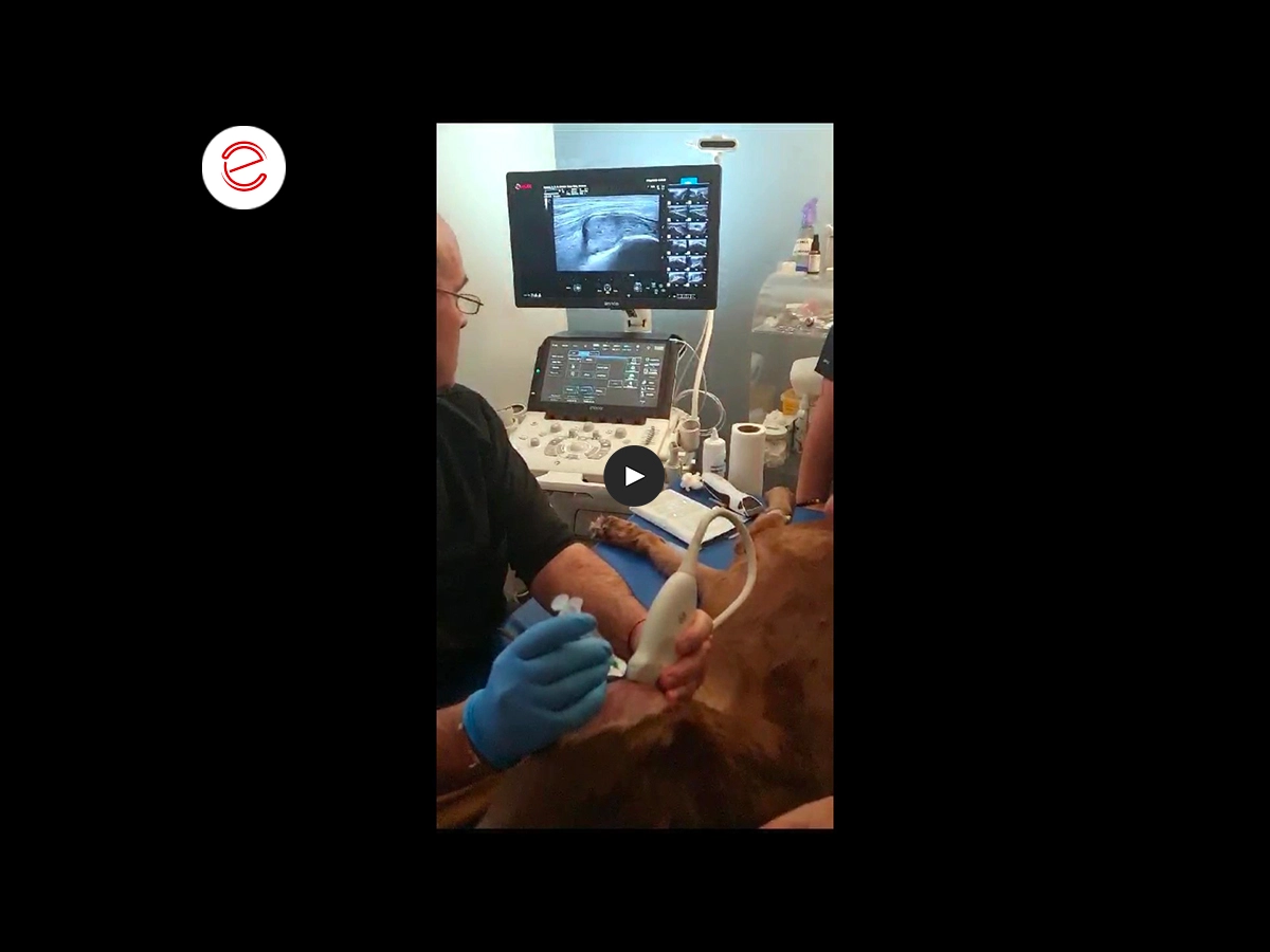
An ultrasound examination of the knee was performed.
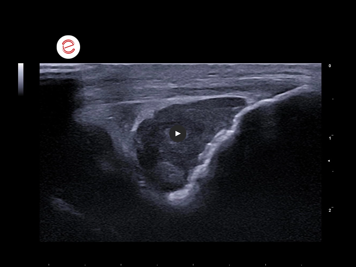
During examination of the joint in the cranial sagittal plane, slightly heterogeneous hypoechoic tissue with a pleomorphic appearance was observed in the infrapatellar adipose body region.
During the lateral sagittal plane examination, the tissue surrounding the tendon of the long digital extensor muscle appears to be the same tissue that projects distally toward its bursa.
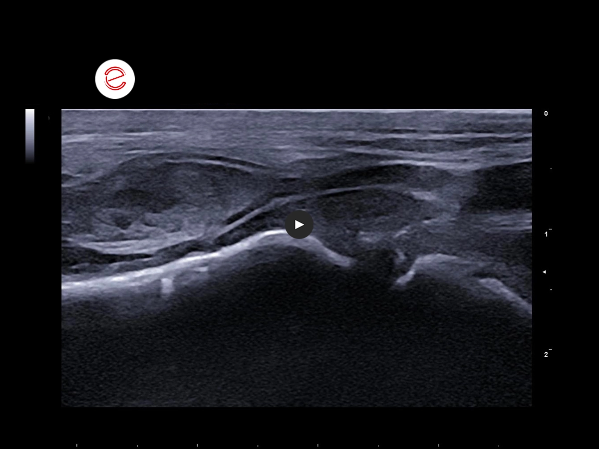
During transverse examination of the area of the femoral trochlea, the same tissue is seen occupying the joint space and protruding proximally toward the supratrochlear bursa.
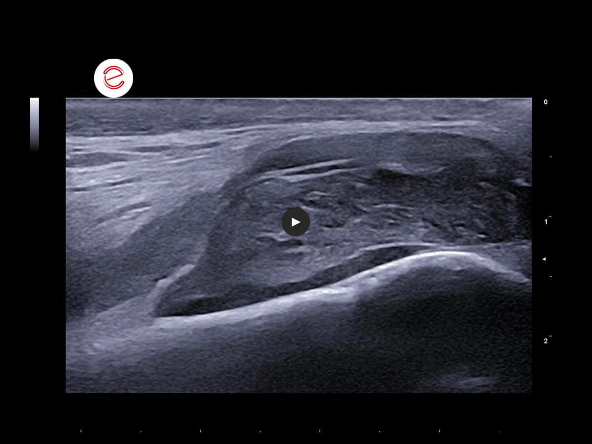
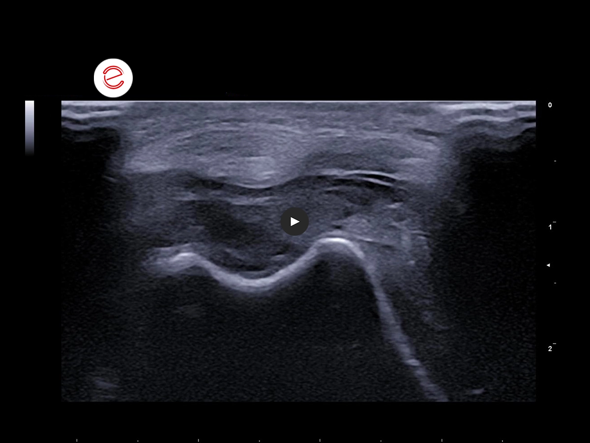
In the sagittal plane above the supratrochlear bursa, the abnormal tissue is seen filling the bursa completely.
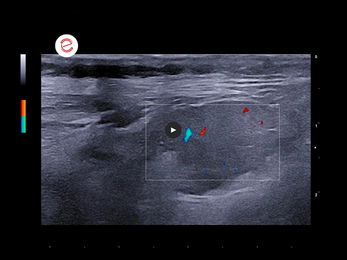
Doppler evaluation of tissue showed mild perfusion.
These findings led to the suspicion of an intra-articular neoproliferative process. Orthogonal knee X-rays and a chest X-ray analysis were performed, as well as a fine-needle aspiration of the abnormal tissue.
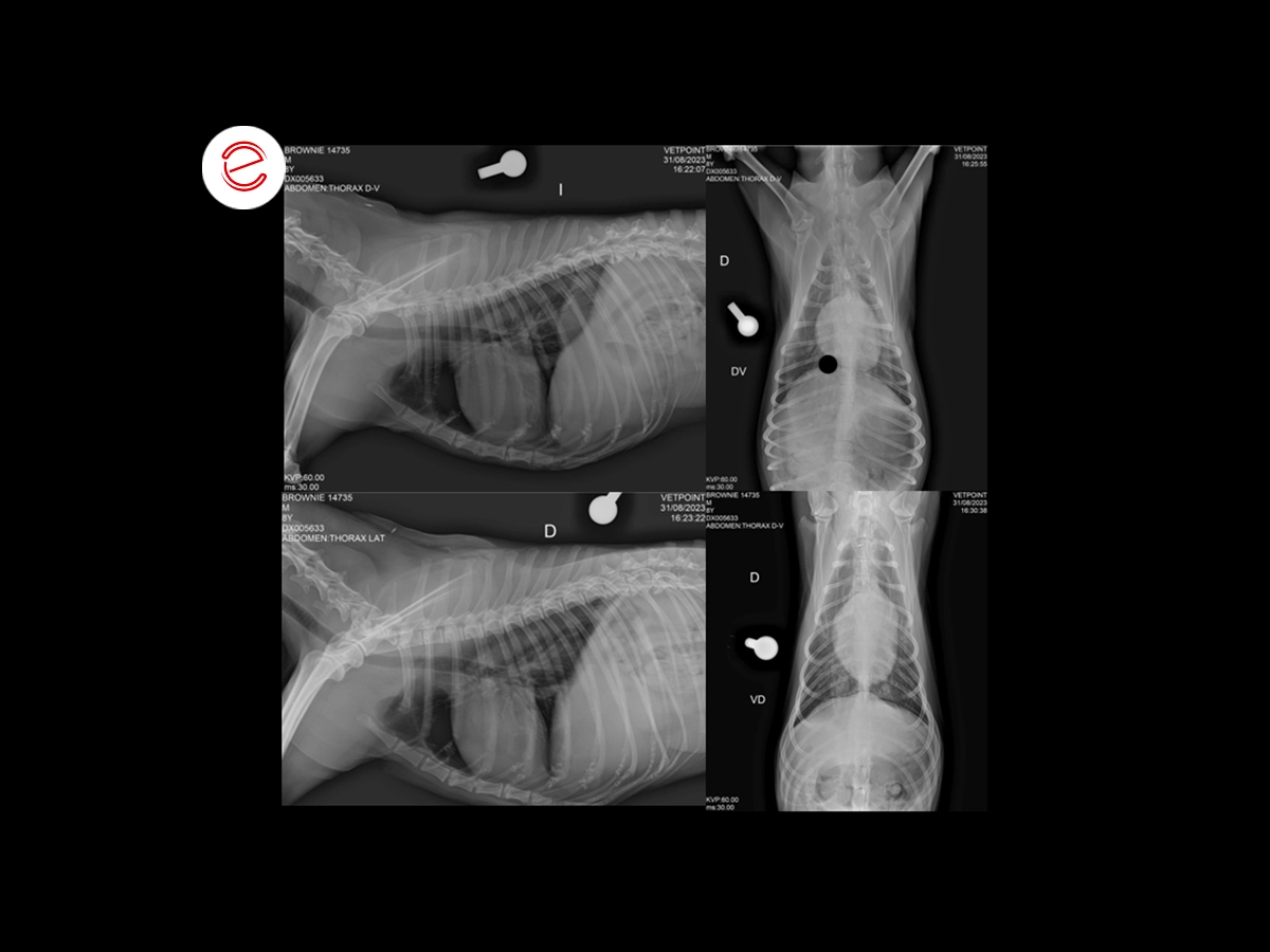
No pathological findings were found during the four-view chest X-ray analysis.
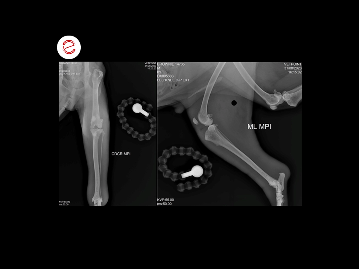
The orthogonal projection of the knee showed osteolysis foci in a moth-eaten pattern in the proximal tibial epiphysis with moderate cortical involvement and no evident periosteal reaction. The infrapatellar adipose body showed an area of tissue density that projected towards the supratrochlear bursa, causing the patella to separate from the femoral trochlea. An aggressive tibial lesion associated with intra-articular tissue/fluid was noted.
A fine-needle tissue aspiration was performed in the area of the supratrochlear bursa.
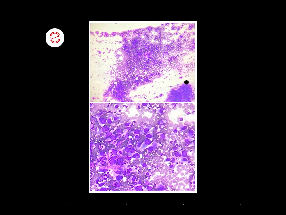
The specimen is moderately cellular with slight hemodilution. Large individualized cells are present with marked anisocytosis and anisokaryosis, and a variable N:C ratio. The cells have an oval to fusiform shape with basophilic cytoplasm and an oval nucleus with finely punctate chromatin, and prominent nucleoli are often seen.
Images were acquired using the MyLab™X90VET system.
Conclusions and treatment
A synovial sarcoma was diagnosed and the limb was amputated.
Daniel Sáez, DVM
Centro de Diagnóstico Veterinario Vetpoint®, Chile

MyLab is a trademark of Esaote spa.
Technology and features are device/configuration-dependent. Specifications subject to change without notice. Information might refer to products or modalities not yet approved in all countries. Product images are for illustrative purposes only. For further details, please contact your Esaote sales representative.
Other canine clinical cases you may be interested in
Discover the challenges faced, the examinations performed, the solutions adopted, and the treatments recommended.

JANUARY 2026
Mammary intraductal carcinoma
Chiara Donà, DVM, PhD, Faculty of Veterinary Medicine, University of Milan, Italy

NOVEMBER 2025
Myxomatous mitral valve disease and heartworm infection
Paul A. Cardenio DVM, MS, DPCCP, DPCVS-CA, Medical Director, PetLandia Veterinary Clinic, City of Malolos, Bulacan, Philippines

JULY 2025
Fibroliposarcoma and malignant melanoma
Reynaldo Buan Jr., DVM, MSc - Buan Veterinary Practice Pampanga, Filippine
